
What Is a Type 2 Polyp? NICE Classification Explained
Author: Dr Sagar Rajkuwar, ENT Specialist, Nashik, Maharashtra, India
🌐 www.entspecialistinnashik.com
Based on the NICE (Narrow-Band Imaging International Colorectal Endoscopic) categorization, a Type 2 polyp is an adenomatous polyp (adenoma) that is very likely to be precancerous. These polyps have irregular vessels and surface patterns that set them apart from non-malignant Type 1 hyperplastic polyps, and they seem to be a darker brown color than the surrounding tissue.
Type 2 Polyps: Important Features
Pathology:
They are often adenomas (tubular, tubulovillous, or villous), which are pre-cancerous, meaning they have the potential to develop into cancer over time.
NICE Type 2 Appearance:
Color: Browner or darker than the neighboring normal tissue.
Brown, thick vessels surround a white, central region.
Surface Pattern: Oval, tubular, or branched structures.
Management:
Due to their neoplastic nature, or capacity to become malignant, Type 2 polyps are often removed during a colonoscopy.
Distinction:
They are distinct from Type 1 (hyperplastic/benign) and Type 3 (suspected deep submucosal cancer) polyps.
Furthermore Subgrouping
The JNET classification system, which is more thorough and similar, is used by some professionals. It distinguishes between Type 2A lesions, which are regular structures, and Type 2B lesions, which are irregular structures and frequently suggest a higher degree of dysplasia or superficial invasion.
Table of contents
- NICE Classification of Colon Polyps: Types, Features, and Clinical Importance
- Types of NICE Classification
- NICE Classification Table
- The NICE Classification’s Benefits
- Clinical Significance in Colon Cancer Screening
- The Nice Classification’s shortcomings
- Conclusion
- FAQ: What Is a Type 2 Polyp?
NICE Classification of Colon Polyps: Types, Features, and Clinical Importance
The NICE Classification, also known as the NBI International Colorectal Endoscopic Classification, is a popular endoscopic method that uses narrow-band imaging (NBI) during colonoscopy to assess and categorize colon polyps. Based on color, vascular patterns, and surface features, this classification assists physicians in distinguishing between hyperplastic polyps, adenomas, and malignant colorectal lesions.
Particularly helpful for evaluating tiny colorectal polyps that are less than 5 or 10 mm in diameter is the NICE categorization. It aids gastroenterologists in determining the optimum course of therapy, such as whether to monitor or remove a polyp, and it enhances real-time diagnosis during colonoscopy.
What is the NICE classification?
The NICE classification was created to make optical diagnosis easier during colonoscopy using narrow-band imaging technology. The same image-enhanced diagnostic methods are available with equipment from other manufacturers, albeit closely related to Olympus Corporation endoscopes.
Three primary visual factors are used in this classification:
- The lesion’s hue
- vascular arrangement
- Surface design
Lesions are categorized into three classes depending on these characteristics.
Types of NICE Classification
Type 1 — Hyperplastic Polyp
Typically, Type 1 lesions are benign hyperplastic polyps that have a very low chance of becoming malignant.
Important Features
Color
- The same color as the surrounding mucosa
- May be lighter than the surrounding tissue at times
Ships
- Only a few or none of the blood vessels are seen.
- limited vascular network
- Lack of an organized vascular pattern
Surface Pattern
- consistent white or dark patches
- Small dots arranged in a circle
- The center is darker, while the surrounding mucosa is lighter.
The Pathology is Most Likely
- Hyperplastic polyp
Relevance to Clinical Practice
In general, hyperplastic polyps are thought to be noncancerous and do not usually need intensive therapy.
Type 2 — Adenoma
Adenomatous polyps, which have the potential to develop into cancer, are known to have type 2 lesions.
Main Characteristics
Color
- More brown than the surrounding mucosa
Ships
- Brown boats encircling lighter buildings
- More noticeable vascular pattern
Surface Pattern
- tubular or oval forms
- A branched or gyrate appearance
- Brown vessels around white core structures
Pathology That Is Most Likely
- Adenoma
- Dysplasia of a lower grade
- Dysplasia of a high grade
- Surface submucosal cancer
Importance in the Clinic
If left untreated, adenomatous polyps may develop into colorectal cancer. The possibility of developing cancer is greatly reduced by early identification and treatment.
Type 3 – Invasive Cancer of the Deep Submucosa
Type 3 lesions are highly related to aggressive colorectal cancer that spreads to deeper layers.
Essential Features
Color
- in comparison to the surrounding tissue, it is a dark brown color.
- Occasionally, there are areas of white that are patchy.
Vessels
- Vessels that are lost or interrupted
- abnormal vascular structure
Surface Design
- Without a clear shape
- Lack of surface structure
The Most Probable Pathology
- Invasive cancer of the deep submucosa
Relevance to Clinical Practice
Due to their high risk of invasive cancer, type 3 lesions frequently need surgical treatment.
NICE Classification Table
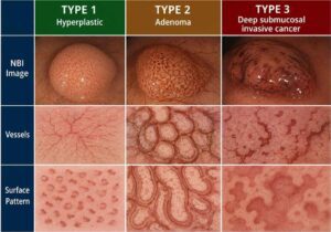
| NICE Type | Color | Vessels | Surface Pattern | Most Likely Diagnosis |
|---|---|---|---|---|
| Type 1 | Same or lighter than background | Sparse or absent vessels | Uniform spots or absent pattern | Hyperplastic polyp |
| Type 2 | Brown relative to background | Brown vessels around white structures | Oval, tubular, or branched structures | Adenoma |
| Type 3 | Dark brown with patchy white areas | Disrupted or absent vessels | Amorphous or absent pattern | Deep invasive cancer |
The NICE Classification’s Benefits
Optical Diagnosis in Real Time
During a colonoscopy, physicians may evaluate polyp features right away without having to wait for pathology results.
Enhanced Identification of Precancerous Lesions
By categorizing adenomatous polyps, it is possible to detect them early before they turn cancerous.
Improved Treatment Choices
With an endoscope, it is possible to ascertain if lesions need:
- polypectomy
- Endoscopic mucosal removal
- Surgery
- Surveillance
Usable With or Without Enhancement
With regular or magnifying colonoscopes, the NICE classification is compatible.
Clinical Significance in Colon Cancer Screening
One of the most prevalent gastrointestinal cancers in the world is colorectal cancer. Colonoscopy’s early detection of adenomas and malignant lesions greatly raises survival rates.
The NICE classification promotes:
- Finding cancer early
- Decreasing the number of unnecessary biopsies
- Improved accuracy of endoscopy
- Enhanced methods for preventing colorectal cancer
The Nice Classification’s shortcomings
Despite its great utility, the NICE system has certain shortcomings:
- The endoscopist’s experience determines how accurate the procedure is.
- Adenomas can resemble inflammatory lesions.
- Histopathology is necessary to confirm certain lesions.
- Additional imaging may still be required for a thorough evaluation of the invasion.
Conclusion
Using narrow-band imaging based on color, vascular structure, and surface patterns, the NICE Classification is a crucial tool in contemporary colonoscopy for categorizing colon polyps. Type 1 lesions are typically hyperplastic, whereas Type 2 lesions often indicate adenomas, and Type 3 lesions provide a strong indication of invasive colorectal cancer.
This classification system facilitates earlier identification of precancerous and cancerous colorectal lesions, enabling physicians to provide timely and successful therapy. The NICE classification continues to be a significant tool in enhancing diagnostic precision during endoscopic assessment as colon cancer screening becomes more and more crucial.
FAQ: What Is a Type 2 Polyp?
What is a Type 2 polyp in the NICE classification?
A Type 2 polyp in the NICE Classification usually refers to an adenomatous colon polyp, which is considered a precancerous lesion with the potential to develop into colorectal cancer over time.
What does a Type 2 colon polyp look like?
Type 2 polyps typically appear browner than the surrounding tissue and show visible brown blood vessels surrounding white oval or tubular surface structures during narrow-band imaging colonoscopy.
Is a Type 2 polyp cancerous?
Most Type 2 polyps are not cancerous, but they may contain low-grade or high-grade dysplasia, meaning abnormal cells that can increase cancer risk if left untreated.
Are Type 2 polyps dangerous?
Type 2 adenomatous polyps can become dangerous over time because some may progress to Colorectal Cancer if not removed during colonoscopy.
How are Type 2 polyps treated?
Doctors usually remove Type 2 polyps during colonoscopy using procedures such as polypectomy or endoscopic mucosal resection.
Can Type 2 polyps come back?
Yes, new adenomatous polyps may develop in the future, which is why regular colonoscopy follow-up is important.
What causes Type 2 adenoma polyps?
Risk factors include:
- Aging
- Family history of colon cancer
- Smoking
- Obesity
- Low-fiber diet
- Inflammatory bowel disease
Are all Type 2 lesions adenomas?
Most Type 2 lesions represent adenomas, including lesions with low-grade dysplasia, high-grade dysplasia, or superficial submucosal carcinoma.
How is a Type 2 polyp diagnosed?
Type 2 polyps are diagnosed during colonoscopy using narrow-band imaging technology and confirmed through biopsy or histopathology.
Can Type 2 polyps be prevented?
Healthy lifestyle habits, regular screening colonoscopy, a high-fiber diet, exercise, and avoiding smoking may help reduce the risk of adenomatous polyps.
Medical Disclaimer
This content is for educational and informational purposes only and should not be considered medical advice, diagnosis, or treatment. Information about Type 2 polyps and the NICE Classification is intended to improve general understanding of colonoscopy findings. Always consult a qualified gastroenterologist or healthcare professional for proper evaluation, diagnosis, and treatment recommendations.
Do not ignore professional medical advice or delay seeking care because of information read online. If you experience symptoms such as rectal bleeding, unexplained weight loss, abdominal pain, or changes in bowel habits, seek medical attention promptly.
👉 YouTube Channel:
http://www.youtube.com/@healthuseful8539/
📞 ENT Consultation & Surgery
Dr. Sagar Rajkuwar (MS-ENT)
Prabha ENT Clinic, Ambad, Nashik
📱 7387590194 | 9892596635
🌐 www.entspecialistinnashik.com
📲 For Health-Related Articles & Videos
🔹 Facebook: https://www.facebook.com/positivemind.healthcare
🔹 YouTube: http://www.youtube.com/@healthuseful8539/
🔗 Related Articles on Nasal Polyps (ENT Expert Guides)
For detailed, evidence-based information, explore the following articles:
https://healthuseful.com/can-nasal-polyps-turn-cancerous/
https://healthuseful.com/can-nasal-polyps-affect-the-brain/
https://healthuseful.com/what-are-the-symptoms-of-cancerous-nasal-polyps/
https://healthuseful.com/when-to-worry-about-nasal-polyp/
https://healthuseful.com/what-happens-if-nasal-polyps-untreated/
https://healthuseful.com/number-one-cause-of-nasal-polyps/
https://healthuseful.com/how-to-stop-polyp-growth/
https://healthuseful.com/can-nasal-polyps-cause-heart-problems/
https://healthuseful.com/how-to-cure-nasal-polyps-permanently/
https://healthuseful.com/can-ent-see-nasal-polyps/
🔗 Sinus Related Articles (Internal Links)
🤧 Sinus Headache & Symptoms
- https://healthuseful.com/sinus-headache-symptoms/
- https://healthuseful.com/sinus-headache-symptoms-one-side/
- https://healthuseful.com/sinus-headache-symptoms-right-side/
- https://healthuseful.com/sinus-headache-back-of-head/
- https://healthuseful.com/sinus-headache-symptoms-home-remedies/
- https://healthuseful.com/how-to-rid-of-a-sinus-infection-headache/
🦠 Sinus Infection Treatment
- https://healthuseful.com/how-to-get-rid-of-a-sinus-infection-in-24-hours/
- https://healthuseful.com/how-to-get-rid-of-sinus-infection-without-antibiotics/
- https://healthuseful.com/what-kills-a-sinus-infection-naturally/
- https://healthuseful.com/sinus-headache-tablet/
- https://healthuseful.com/how-do-doctors-drain-sinuses-without-surgery/
- https://healthuseful.com/signs-of-sinus-infection-getting-better/
- https://healthuseful.com/signs-of-sinus-infection-getting-better-with-antibiotics/
⚠️ Serious Conditions & Warning Signs
- https://healthuseful.com/end-stages-of-a-sinus-infection/
- https://healthuseful.com/how-to-tell-if-sinus-infection-has-spread-to-brain/
- https://healthuseful.com/how-to-tell-if-sinus-infection-has-spread-to-bone/
- https://healthuseful.com/what-is-a-red-flag-for-sinusitis/
- https://healthuseful.com/when-to-go-to-ae-with-sinusitis/
👁️ Related Symptoms & Complications
- https://healthuseful.com/can-a-sinus-infection-cause-eye-pain/
- https://healthuseful.com/can-sinusitis-affect-your-ears/
- https://healthuseful.com/can-sinus-infection-cause-hair-loss/
- https://healthuseful.com/symptoms-of-perforated-sinus-after-tooth-extraction/
🧠 Causes, Triggers & Misdiagnosis
- https://healthuseful.com/what-triggers-sinusitis-headaches/
- https://healthuseful.com/what-can-be-mistaken-for-a-sinus-infection/
- https://healthuseful.com/what-mimics-sinus-issues/
- https://healthuseful.com/what-can-worsen-a-sinus-infection/
🧬 Nutrition & Deficiency
- https://healthuseful.com/what-vitamin-deficiency-causes-sinus-headaches/
- https://healthuseful.com/what-vitamin-deficiency-causes-sinusitis/
- https://healthuseful.com/what-deficiency-causes-sinusitis/
📘 General Sinus Knowledge
- https://healthuseful.com/what-are-the-four-cardinal-signs-of-sinusitis/
- https://healthuseful.com/is-a-sinus-infection-contagious-with-fever/
- https://healthuseful.com/can-you-smell-a-fungal-sinus-infection/
- https://healthuseful.com/sinusitis-in-hindi/
💰 Treatment & Surgery
- https://healthuseful.com/cost-of-sinus-surgery-in-india/
- https://healthuseful.com/how-to-cure-sinus-permanently/
🧒 Special Cases
🔍 Rare Conditions
References
- American Society for Gastrointestinal Endoscopy (ASGE) – Guidelines and educational resources on colonoscopy and colorectal polyp classification.
- World Endoscopy Organization – Information on advanced endoscopic imaging and colorectal lesion assessment.
- National Cancer Institute – Educational material about colorectal polyps and colorectal cancer screening.
- Mayo Clinic – Medical overview of colon polyps, symptoms, and treatment options.
- Cleveland Clinic – Clinical information about adenomatous polyps and colonoscopy findings.
- PubMed – Research database containing studies on the NICE classification and narrow-band imaging.
- American Cancer Society – Resources on colorectal cancer prevention and colon polyp detection.


