Cholesteatoma Complications: Symptoms, Causes, Diagnosis and Treatment
Cholesteatoma can lead to serious complications including hearing loss, facial nerve damage, and life-threatening infections if left untreated.
Author: Dr. Sagar Rajkuwar (MS-ENT), ENT Specialist, Nashik, Maharashtra, India
Clinic: Prabha ENT Clinic, Ambad, Nashik
🌐 www.entspecialistinnashik.com
Updated October 2026 with the latest ENT evidence on intracranial complication rates in the modern antibiotic era, facial-nerve palsy outcomes and timing of decompression, labyrinthine-fistula management, pediatric vs adult cholesteatoma behavior, lateral sinus thrombosis treatment, and non-echo-planar DWI MRI surveillance.
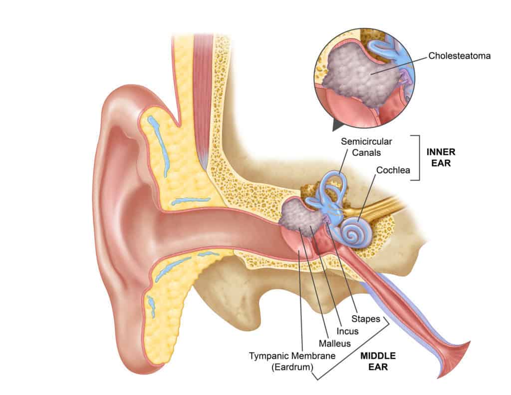
Figure 1 — Cholesteatoma complications: how disease spreads from the temporal bone to intracranial structures.
Table of Contents
- What Is Cholesteatoma?
- What Causes Cholesteatoma?
- What Are the Symptoms of Cholesteatoma?
- How Does Cholesteatoma Cause Complications?
- Cholesteatoma Complications
- How Is Cholesteatoma Diagnosed?
- When Is Cholesteatoma an Emergency?
- Treatment of Cholesteatoma
- Tympanomastoidectomy for Cholesteatoma
- Canal Wall-Up vs Canal Wall-Down Surgery
- Can Medicines Cure Cholesteatoma?
- Recovery and Follow-Up After Surgery
- Can Cholesteatoma Come Back?
- Frequently Asked Questions
- Modern Advances in Cholesteatoma Management (new, Oct 2026)
- Summary
1. What is Cholesteatoma?
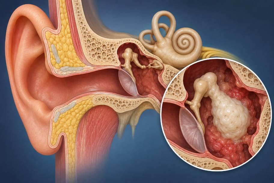
The middle ear and, potentially, the mastoid bone are often affected by a cholesteatoma, an uncommon buildup of keratinizing skin cells and debris.
A cholesteatoma is not a malignant tumor, despite its name. Nonetheless, it may behave destructively because it can expand and wear away the sensitive tissues and bone around the ear.

Cholesteatoma might be:
- Congenital, meaning it is there from birth.
- later in life.
Eustachian tube dysfunction, tympanic membrane retraction pockets, chronic ear infection, trauma, and prior ear surgery are all often linked to acquired cholesteatoma.
Further context (added October 2026): Beyond the simple congenital/acquired split, modern otology recognises four clinically useful categories: (i) congenital cholesteatoma, forming behind an intact drum and often found incidentally in children; (ii) primary acquired cholesteatoma, arising from a pars-flaccida or pars-tensa retraction pocket in a continuously under-ventilated middle ear; (iii) secondary acquired cholesteatoma, forming after a traumatic or infected perforation when squamous epithelium migrates in from the external canal; and (iv) iatrogenic cholesteatoma, from implanted skin after previous ear surgery. Each category tends to behave slightly differently — primary acquired disease often spreads first into the attic and antrum, while congenital disease classically starts in the anterior mesotympanum. Published European and NHS estimates place the overall incidence of acquired cholesteatoma at roughly 9–12 new cases per 100,000 adults per year and 3–6 per 100,000 children per year, with higher rates in populations with poor Eustachian-tube function, cleft palate, Down syndrome, or recurrent childhood otitis media.
2. What is the cause of cholesteatoma?
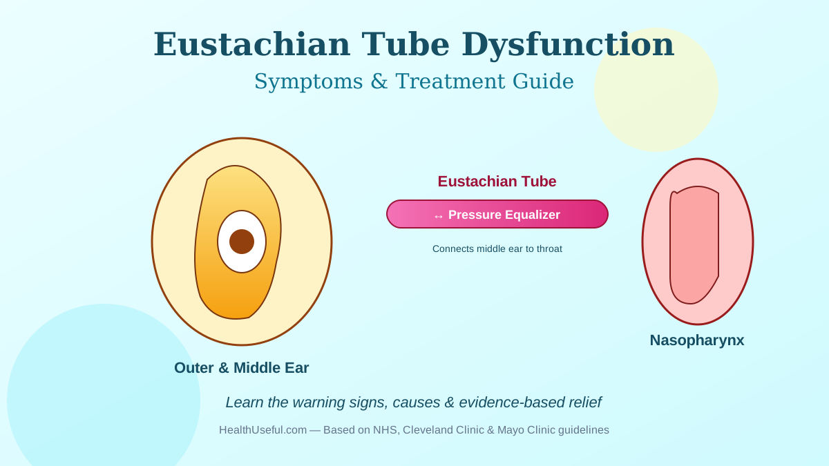
Poor ventilation of the middle ear is a frequent method.
When the eustachian tube isn’t working as it should, there can be a buildup of negative pressure in the middle ear. Part of the eardrum can be pulled inward by this, forming a retraction pocket.
This cavity might collect dead skin cells. The collection may grow and become a cholesteatoma over time.
Other possible causes include:
- persistent otitis media
- Perforation of the eardrum
- Prior ear injury
- Prior Ear Operation
- Abnormalities present at birth
The precise process can differ from one patient to another.
Further context (added October 2026): There are four main scientific theories for how acquired cholesteatoma develops, and they are not mutually exclusive: (1) retraction-pocket theory — chronic Eustachian-tube dysfunction creates persistent negative middle-ear pressure, the pars flaccida retracts, keratin stops self-cleaning and accumulates; (2) migration theory — squamous epithelium from the external canal invades the middle ear through a marginal perforation; (3) metaplasia theory — the respiratory mucosa of the middle ear transforms into squamous epithelium under chronic inflammation; and (4) basal-cell hyperplasia / papillary ingrowth theory — the basal layer of the pars flaccida sends down fingers of keratinising epithelium that enlarge over time. Risk factors identified in the latest literature include recurrent acute otitis media in childhood, cleft palate and craniofacial syndromes, Down syndrome, allergic rhinitis with chronic Eustachian-tube obstruction, previous ventilation-tube insertion, passive tobacco-smoke exposure, and biofilm-forming middle-ear infection. Understanding the specific mechanism in a given patient matters because it guides prevention of recurrence after surgery — a patient with persistent Eustachian-tube dysfunction, for example, benefits from medical management of the nose and nasopharynx (allergy treatment, adenoidectomy where indicated, nasal steroids) alongside the ear surgery itself.
3. What are the symptoms of cholesteatoma?
Symptoms of cholesteatoma are occasionally missed and it might develop gradually.
Typical symptoms include:
- Frequent or persistent ear discharge
- Foul-smelling discharge
- Slow hearing loss
- Fullness in the ear
- A sensation of discomfort or pain in the ear.
- Tinnitus
- Vertigo or dizziness
- Chronic ear infections
An ENT professional should evaluate a chronic, foul-smelling ear discharge that doesn’t significantly improve with standard care.
Added October 2026 — symptom character and red-flag combinations: The discharge in cholesteatoma is classically scanty, thick, cheesy or pearly, and has a distinct foul odor from anaerobic bacterial growth within trapped keratin. It differs from the thin, pus-like discharge of ordinary chronic suppurative otitis media. Important red-flag combinations that should prompt same-day ENT evaluation include: discharge PLUS new facial weakness (facial-nerve involvement); discharge PLUS vertigo brought on by pressure on the tragus (positive fistula sign suggesting labyrinthine erosion); discharge PLUS severe headache, high fever or neck stiffness (suspected meningitis or intracranial abscess); discharge PLUS sudden severe hearing loss (labyrinthitis or inner-ear involvement); and discharge PLUS swelling behind the ear with the pinna pushed outwards (acute mastoiditis with possible abscess). Congenital cholesteatoma in children is often painless and detected only on routine ear examination as a white pearl behind an intact eardrum, which is why schools and paediatricians increasingly include otoscopy in standard childhood check-ups.
4. What are the mechanisms of cholesteatoma complications?
Bone erosion can occur as a result of the slow growth of a cholesteatoma.
The affected structures could be:
- Bones, little ones.
- Canal of the facial nerve
- Structures of the inner ear
- The mastoid bone
- Middle ear or mastoid tegment
- Sigmoid sinus-enveloping structures
In patients with advanced illness, the infection or erosion might spread to the interior of the skull.
Because of this, a cholesteatoma should not be seen as a basic ear infection.
Added October 2026 — the biology behind the destruction: Cholesteatoma does not simply push bone aside — it actively destroys it. The expanding keratin mass releases pro-inflammatory cytokines (TNF-α, IL-1, IL-6), prostaglandins and matrix metalloproteinases that stimulate osteoclast activity along the margin of the lesion, so bone is actively resorbed, not just compressed. Research has also identified RANKL/OPG signalling as a central driver of this osteoclast activation, and biofilms of Pseudomonas aeruginosa and Staphylococcus aureus on the keratin surface amplify the inflammatory signal. This is why antibiotics alone cannot cure the disease — the mechanical mass and its biochemical environment remain even when overlying infection is suppressed, and the bone destruction continues silently. The sequence of structures affected depends on the location of the pocket: pars-flaccida (attic) cholesteatomas tend to erode the scutum first, then the long process of the incus and the head of the malleus, and may then reach the lateral semicircular canal superiorly; pars-tensa cholesteatomas often reach the stapes superstructure and oval window early; and advanced disease in either location can reach the tegmen tympani, sigmoid sinus plate, facial canal and labyrinth.
5. Complications of Cholesteatoma
5.1 Hearing Loss
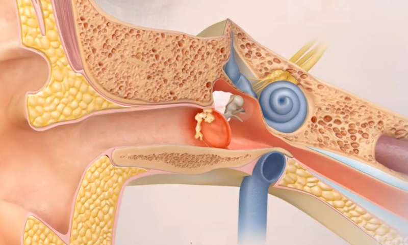
Among the most frequent consequences of cholesteatoma is hearing impairment.
The cholesteatoma might harm the ossicles, which are small bones that send sound through the middle ear.
This frequently results in conductive hearing loss.
Mixed hearing loss may result if the patient’s inner ear is also damaged, causing a sensorineural component.
The degree of hearing loss is determined by the area and scope of the illness.
Added October 2026 — numbers that help patients understand their own situation: Published series describe conductive hearing loss in roughly 60–90% of patients at diagnosis, with air–bone gaps typically between 20 and 50 dB depending on how many ossicles are involved. The long process of the incus is the single most commonly eroded ossicle (reported in up to 70% of surgical specimens), followed by the stapes superstructure, then the malleus head. Sensorineural components are reported in 10–30% of advanced cases, usually from labyrinth erosion or inner-ear toxin diffusion through the oval or round window. Ossicular reconstruction (titanium or hydroxyapatite partial or total ossicular replacement prostheses — PORP/TORP) at the time of surgery can close the air–bone gap to within 20 dB in approximately 60–70% of patients when disease has been fully cleared and the inner ear is intact.
5.2 Facial Palsy or Facial Nerve Weakness
Near the middle ear, the temporal bone is penetrated by the facial nerve.
Inflammation and compression can result from a cholesteatoma’s erosion of the facial nerve’s bony covering.
This can lead to:
- weakness in the face
- asymmetry in the face
- Trouble shutting one eye
- One side of the face sags.
Facial weakness is a serious warning indicator that necessitates quick ENT evaluation.
Added October 2026 — incidence, timing and recovery: Facial nerve palsy complicates cholesteatoma in roughly 1–3% of cases in modern series, rising to 5–8% in patients from low-resource settings presenting late. In published case series (including PMC9844432 and similar cohorts), the tympanic (horizontal) segment of the facial nerve is the single most commonly involved site because its bony canal is naturally dehiscent in up to 30% of normal temporal bones. Mechanisms are compression by the keratin mass, infective neuritis, and direct erosion of the facial canal. The strongest predictor of recovery is time to surgical decompression: patients operated within 2–3 weeks of onset of palsy achieve House–Brackmann grade I–II recovery in roughly 70–80% of cases, while recovery drops to around 30–40% when surgery is delayed beyond 3 months. Modern practice therefore treats a new facial palsy in cholesteatoma as a surgical emergency: urgent high-resolution CT of the temporal bone, intravenous antibiotics (usually a third-generation cephalosporin plus metronidazole where anaerobes are likely), systemic corticosteroids, and tympanomastoidectomy with facial-nerve decompression within days rather than weeks.
5.3 Inner ear damage and vertigo
A cholesteatoma can eat away at structures near the inner ear.
Erosion of the labyrinth, especially, can cause a labyrinthine fistula or other inner ear damage.
Symptoms might include:
- Dizziness
- Feeling lightheaded
- Problems with balance
- Vomiting
- Hearing loss of the sensorineural type
Therefore, vertigo in a patient with known or suspected cholesteatoma must be properly evaluated.
Added October 2026 — labyrinthine fistula in detail: Labyrinthine fistula — a bony defect in the labyrinth exposing the membranous inner ear — is reported in 4–12% of cholesteatoma surgeries, with the lateral (horizontal) semicircular canal involved in approximately 90% of fistulas. Classic clinical findings include Hennebert’s sign (vertigo and nystagmus triggered by pressure applied to the external canal) and Tullio’s phenomenon (vertigo triggered by loud sounds). High-resolution CT of the temporal bone identifies most fistulas preoperatively. Surgical management is nuanced: the overlying matrix is sometimes left in situ over the fistula to preserve inner-ear function, with careful postoperative follow-up and non-EPI DWI MRI surveillance, because aggressive removal can cause total sensorineural hearing loss (“dead ear”) in roughly 10–25% of cases. Specialist otology units now routinely preserve hearing in the majority of fistula patients using modern microsurgical and endoscopic techniques, bone-pâté sealing, and in selected cases staged surgery.
5.4 Chronic Foul-Smelling Discharge and Ear Infection
A persistent or recurring ear infection is commonly caused by cholesteatoma.
Keratin debris and dead skin might build up inside the lesion, promoting ongoing inflammation and infection.
Patients might observe:
- Ongoing ear discharge
- offensive odor
- Recurring infections
- Pain in the ears
- Diminishment of hearing
While antibiotic ear drops might help control any related infection, they won’t get rid of the cholesteatoma that’s causing it.
Added October 2026 — microbiology and biofilm: The dominant organisms isolated from infected cholesteatoma are Pseudomonas aeruginosa, Staphylococcus aureus, and mixed anaerobes (including Bacteroides and Prevotella species) which produce the characteristic foul odor. Up to 80% of these organisms form biofilms on the keratin surface, which blunt the effect of ordinary ear drops and allow infection to relapse as soon as treatment stops — one more reason that medical treatment alone cannot cure cholesteatoma. Evidence-based topical therapy for a flare-up is a topical fluoroquinolone (ciprofloxacin, or ciprofloxacin + dexamethasone where available) because it covers the common pathogens, is non-ototoxic even if the middle ear is exposed, and penetrates biofilm better than older agents. Aminoglycoside drops (gentamicin, neomycin) in an exposed middle ear should be avoided outside specialist guidance because of potential sensorineural ototoxicity.
5.5 Tinnitus
In certain individuals with cholesteatoma, tinnitus can manifest as ringing, buzzing, humming, or other noises in the afflicted ear.
Tinnitus may be present with hearing loss or involvement of the middle or inner ear.
Added October 2026: Tinnitus accompanies cholesteatoma in roughly 40–50% of patients at diagnosis and typically improves after successful disease clearance, provided the sensorineural system has not been damaged. Pulsatile tinnitus in a patient with cholesteatoma is a specific red flag — it may reflect sigmoid-sinus plate erosion, developing lateral sinus thrombosis, or an abnormal vascular communication, and warrants same-day imaging (CT with contrast or MR venogram).
5.6. Erosion of the temporal bone and ossicles
The tiny bones of hearing and other sections of the temporal bone might be corroded by the cholesteatoma.
Significant hearing loss can result from ossicular damage.
More widespread disease can include:
- Scutum
- Air cells of the mastoid bone
- Tegmen
- Canal of the facial nerve
- Labyrinth
- Additional vital structures of the temporal bone
Cholesteatoma has been shown to erode the jugular bulb, intracranial structures, tegmen, and sigmoid sinus.
Added October 2026 — frequency of bony involvement in surgical series: Published surgical series (including large Indian, Turkish and European cohorts) report the following approximate frequencies of bone erosion at the time of surgery: scutum 60–80%; incus long process 55–70%; stapes superstructure 25–40%; mastoid tegmen 15–25%; facial canal (any segment, often naturally dehiscent) 30–50%; lateral semicircular canal 4–12%; sigmoid sinus plate 10–20%; dural plate 10–15%; petrous apex involvement is rare (<2%). The scutum (bony lip over the attic) is almost always eroded in pars-flaccida disease because the pocket itself originates there. The number of structures involved correlates with disease duration — late presentation is the single biggest predictor of extensive erosion.
5.7 Meningitis
Infection brought on by an advanced cholesteatoma can spread toward the meninges and result in meningitis, though it is unusual.
The symptoms may include:
- Intense headache
- Fever
- Stiffness in the neck
- Nausea and vomiting
- Sensitivity to light
- Alterations in consciousness or disorientation
A medical emergency is meningitis.
Added October 2026 — modern incidence and outcomes: Otogenic meningitis is now the single most common intracranial complication of cholesteatoma, accounting for about 40–50% of all intracranial complications. Overall incidence of any intracranial complication in modern series ranges from approximately 1–3% of surgically treated cholesteatoma cases — a sharp fall from the 8–10% rate reported in the pre-antibiotic era — but mortality in those complications remains 5–20% depending on timeliness of diagnosis and surgical intervention. Routes of spread include direct extension through an eroded tegmen, retrograde thrombophlebitis of small veins, and spread along preformed anatomical pathways (dehiscent tegmen tympani, labyrinth, round window). Lumbar puncture is usually deferred until CT excludes abscess (risk of coning), and treatment combines intravenous broad-spectrum antibiotics (meningitic doses crossing the blood–brain barrier), neurosurgical consultation, and urgent ear surgery to drain and remove the source of infection.
5.8 Brain Abscess
Although uncommon, a brain abscess can be fatal.
It can happen when infection from severe ear disease goes beyond the temporal bone and affects intracranial structures.
Among the signs are:
- persistent or severe headache
- Fever
- Vomiting
- Confusion
- Weakness
- Seizures
- Changes in awareness
If a brain abscess is thought to exist, it needs to be examined right away in a hospital.
Epidural abscess and venous sinus thrombosis are additional uncommon intracranial side effects of cholesteatoma.
Added October 2026 — further detail on brain abscess, sinus thrombosis and other intracranial complications:
- Otogenic brain abscess is most often located in the temporal lobe (adjacent to the tegmen) or the cerebellum (adjacent to Trautmann’s triangle), accounts for roughly 15–25% of intracranial otogenic complications, and typically grows from mixed anaerobic and aerobic organisms. Modern management combines neurosurgical aspiration or excision, 6–8 weeks of intravenous antibiotics, and definitive mastoid surgery to remove the source.
- Lateral (sigmoid) sinus thrombosis complicates roughly 10–20% of intracranial otogenic complications. Classic signs include spiking “picket-fence” fever, headache, papilledema, and the Griesinger sign (tender swelling over the mastoid from thrombosis of the mastoid emissary vein). Diagnosis is confirmed by contrast-enhanced CT or MR venography. Modern treatment combines urgent mastoidectomy with exposure of the sinus and removal of necrotic bone over it; anticoagulation is now used in most centres for propagating thrombus or when there is risk of intracranial extension, although it remains debated when thrombus is localised.
- Extradural (epidural) abscess is often discovered during mastoidectomy when infected granulation tissue is found between the tegmen bone and dura; it may be asymptomatic or present with headache alone.
- Subdural empyema is rarer but more dangerous because it spreads rapidly along the subdural space.
- Otitic hydrocephalus — raised intracranial pressure without a mass lesion — is now rarely seen with modern treatment.
- Encephalitis / focal cerebritis can precede a frank abscess and may be reversible with early antibiotics.
Overall, the pattern of intracranial complications has shifted since antibiotics became widely available — abscesses are relatively less common, meningitis and lateral sinus thrombosis relatively more so — but a modern cholesteatoma series that reports zero intracranial complications is rare, and the pattern justifies the ENT consensus that cholesteatoma, once diagnosed, warrants surgical removal in almost every patient fit for anaesthesia.
6. How Is Cholesteatoma Recognized?
A thorough ENT exam typically marks the beginning of the diagnosis.
The doctor might do the following:
Ear Check
You may use an otoscope or an operating microscope to discover:
- Retraction pockets
- Debris from keratin
- Eardrum perforation
- Tissue that granulates
- Unusual discharge
- Test of hearing
Determining the kind and severity of hearing loss is aided by pure-tone audiometry.
CT scan
A high-resolution CT may be used to assess the following:
- Erosion of bone
- Involvement of the mastoid bone
- Damage to the ossicular system
- Anatomy of the face channel
- Additional difficulties
- Diffusion-weighted imaging MRI
Cholesteatoma may be identified, and the residual or recurrent disease following surgery may be assessed using diffusion-weighted MRI, which may be extremely helpful.
Added October 2026 — the role of non-echo-planar DWI MRI: Non-echo-planar diffusion-weighted MRI (non-EPI DWI MRI) is now the imaging modality of choice for suspected residual or recurrent cholesteatoma, and in many European and UK otology units has largely replaced the routine “second-look” operation after canal-wall-up tympanomastoidectomy. The 2022 Cureus systematic review and meta-analysis reported a pooled sensitivity of about 92% and specificity of about 94% for non-EPI DWI MRI in detecting residual/recurrent cholesteatoma, with the smallest reliably detected lesion around 3 mm. Non-EPI DWI MRI is also increasingly used preoperatively, especially in revision cases, to delineate disease extent and plan surgery. Combined with a high-resolution temporal-bone CT, this modern imaging pair gives the surgeon very reliable preoperative information on bone anatomy, erosion, and the location of the keratin mass.
7. When Is Cholesteatoma an Emergency?
Seek immediate medical help if cholesteatoma is accompanied by:
- Recent facial weakness
- Extreme giddiness
- Extreme headache
- Fever accompanied by worsening ear symptoms
- Confusion
- Vomiting repeatedly
- Loss of balance
- Symptoms related to the nervous system
- A sharp and abrupt decrease in hearing capacity.
Intracranial structures, the inner ear, or the facial nerve may be affected by these symptoms.
Added October 2026 — a practical emergency pathway: A patient presenting with any of these red-flag symptoms needs an urgent pathway that includes: (1) same-day ENT examination and microscopy; (2) urgent high-resolution CT of the temporal bones (with contrast if intracranial complication is suspected); (3) MRI with contrast and MR venography when CT raises concern for abscess, cerebritis, or sinus thrombosis; (4) blood cultures and inflammatory markers; (5) urgent neurological or neurosurgical consultation where indicated; (6) empirical intravenous antibiotics (typically a third-generation cephalosporin plus metronidazole, modified by local protocols and allergies); (7) urgent surgical intervention — tympanomastoidectomy with cholesteatoma clearance, often combined with neurosurgical drainage of abscess if present, and with sinus-plate exposure or sinus incision for thrombosis. Delays in this pathway are the single most consistent predictor of poor outcome in the published literature. Patients and families should know that the correct answer to “it is 2 AM, should we go to A&E?” when these symptoms appear is almost always “yes.”
8. Treatment of Cholesteatoma
The kind of therapy used is determined by the size, location, and condition of the patient’s ear, as well as the problems that may be present.
In most cases, surgical removal is the primary form of treatment for a cholesteatoma that has clinical importance.
Medications can be used to manage inflammation or infection, however they are not assured to completely cure the keratinizing disease.
The goals of therapy include:
- Cholesteatoma must be removed.
- Maintain a dry and safe ear.
- Manage infection.
- Protect key structures.
- When feasible, either maintain or enhance hearing.
- Lessen the likelihood of recurrence.
Added October 2026 — modern decision-making and consensus: The European Academy of Otology & Neurotology / Japanese Otological Society joint consensus (updated 2022) formalised cholesteatoma staging using the STAM system — recording involvement of the Supratubal recess, Tympanic cavity, Antrum and Mastoid — plus extensions (E1 difficult sites such as the sinus tympani, E2 fistula, E3 facial nerve, E4 dural exposure, E5 intracranial). This staging now shapes the surgical plan, guides counselling about recurrence risk, and allows meaningful comparison of outcomes between centres. In parallel, modern practice increasingly combines microscope and endoscope (“endoscopic ear surgery”) because the endoscope allows direct visualisation and clearance of difficult hidden recesses — notably the sinus tympani, facial recess and anterior epitympanum — which were previously the main sites of residual disease.
9. Tympanomastoidectomy for Cholesteatoma
When necessary, a tympanomastoidectomy combines mastoid surgery with surgery on the eardrum or middle ear.
Surgery may involve: depending on the illness.
- removal of cholesteatoma
- Mastoidotomy
- Tympanoplasty
- Reconstruction of ossicles
- Removing ailing tissue
- reconstruction of the tympanic membrane
The precise technique is customized based on the disease’s degree.
Consult our thorough manual on cholesteatoma surgery.
Added October 2026 — role of endoscopic ear surgery: Endoscopic ear surgery (EES) has become a mainstream technique for cholesteatoma clearance over the last decade. The endoscope gives direct line-of-sight into hidden recesses (sinus tympani, facial recess, anterior epitympanum, retrotympanum) that the operating microscope reaches only indirectly. Pooled data from EES series suggest residual-disease rates can be lower than with microscope-only surgery, particularly for pars-tensa cholesteatoma and for pediatric cases. EES is often done through an entirely transcanal approach for limited disease, avoiding a post-auricular incision; combined endoscope–microscope approaches are preferred for extensive mastoid involvement. Patients considering surgery for cholesteatoma may ask whether their surgeon uses endoscopic techniques, and whether this would benefit their particular case.
10. Canal wall-up vs canal wall-down surgery
There are two main surgical techniques to choose from.
Up the Canal Wall
The outside ear canal is still intact.
Although this method may result in a more natural anatomy of the ear canal, postoperative monitoring is essential since residual or recurrent illness is possible.
Canal Wall-Down
An open mastoid cavity is created by removing a portion of the posterior wall of the ear canal.
For specific patients with extensive disease or anatomy that necessitates this method for full disease clearance, this may be suitable.
The surgeon’s assessment, prior surgery, hearing condition, anatomy, and extent of the illness are some of the elements that influence the selection.
Added October 2026 — residual and recurrence rates compared: Published pooled series put residual disease rates after canal-wall-up tympanomastoidectomy at approximately 10–25% and after canal-wall-down surgery at 3–10%; recurrence (new disease in a different site) is approximately 5–15% for canal-wall-up and 2–5% for canal-wall-down. Canal-wall-up surgery offers better anatomy for hearing-aid use and for swimming, but requires either a planned second-look operation at 9–18 months or non-EPI DWI MRI surveillance to detect hidden residual disease. Canal-wall-down surgery lowers the risk of hidden disease at the cost of a lifelong mastoid cavity needing periodic professional cleaning. A hybrid approach — canal-wall-up with mastoid obliteration (often using bioactive glass S53P4, bone pâté or cartilage) — is increasingly used by specialist centres because it combines the low recurrence of canal-wall-down with the better anatomy of canal-wall-up.
11. Can Medicines Cure Cholesteatoma?
A cholesteatoma can seldom be eradicated by medication alone.
Systemic antibiotics or antibiotic ear drops may be used if there is accompanying discharge and infection.
However, the underlying keratinizing lesion is not eliminated by medication.
As a result, patients who are thought to have cholesteatoma should be seen by an ENT/otologist rather than just receiving several rounds of antibiotics.
Added October 2026 — role of medical therapy as adjunct, not cure: Medical treatment plays three legitimate roles in the overall management of cholesteatoma: (1) preoperative preparation — a short course of topical fluoroquinolone drops before surgery to dry the ear and reduce infection, which improves surgical visibility and may lower infection-related recurrence; (2) management of flare-ups while awaiting surgery or in patients unfit for operation; and (3) postoperative management of granulation and residual low-grade infection. Experimental medical treatments — topical retinoids, bone-resorption inhibitors, biofilm-dispersing agents — have been investigated but none has yet replaced surgical clearance as the definitive treatment. Patients should be wary of advertised “non-surgical cures” for cholesteatoma: the condition is a mechanical mass of keratin that must be physically removed, and delay allows further bone destruction.
12. Recovery and Follow-Up After Surgery
The procedure performed and the degree of the condition determine how long it takes to recover.
Patients may briefly experience the following after surgery:
- fullness in the ear
- Lower hearing
- A little pain
- tinnitus
- Dizziness
- Taste alteration
Patients should adhere to their surgeon’s recommendations regarding:
- Care for your ears
- Maintaining dry ears
- Drops for ears
- Occupation
- To Fly
- Swimming
- follow-up appointments
Residual illness or the recurrence of cholesteatoma might occur; hence, long-term follow-up is crucial.
Added October 2026 — a typical modern follow-up schedule:
- Week 2–3: wound and ear-canal pack review; suture removal if non-absorbable.
- Week 6–8: microscopic examination, early assessment of graft healing.
- Month 3–6: pure-tone audiogram; consideration of hearing-aid trial if indicated.
- Month 9–12: first surveillance non-EPI DWI MRI (in centres using imaging in place of second-look surgery).
- Year 2–5: annual clinical review; repeat MRI at the surgeon’s discretion. Long-term follow-up is recommended because recurrence can appear up to 10–15 years after apparently successful surgery.
- Patient-reported outcomes are increasingly captured using validated instruments such as COMOT-15, COMQ-12 and ZCMEI-21, which allow comparison of quality-of-life gains across surgical approaches.
Water precautions, cotton-bud avoidance, and prompt reporting of new discharge, hearing change, vertigo or facial weakness are a lifelong part of care in these patients.
13. Can Cholesteatoma Come Back?
Yes.
It’s possible that cholesteatoma will persist or reappear after the procedure.
The risk varies according on a variety of criteria, such as:
- The scope of the initial sickness
- Operative strategy
- The patient’s age.
- Structure
- Whether the illness persisted in areas that were difficult to reach
Long-term clinical and, when necessary, imaging monitoring can aid in the detection of remaining or recurrent disease. Postoperative monitoring is greatly aided by non-echo-planar diffusion-weighted MRI.
Added October 2026 — pediatric vs adult recurrence: Pediatric cholesteatoma is widely accepted to behave more aggressively than adult disease, with recurrence rates roughly 1.5–2× higher. Reasons include a more pneumatised mastoid (more hidden spaces), a thinner matrix that fragments during surgery, more pronounced inflammatory response, and persistent Eustachian-tube dysfunction. Specialist pediatric otology units therefore favour: (a) planned imaging surveillance with non-EPI DWI MRI; (b) endoscopic ear surgery for hidden recesses; (c) staged procedures when clearance is uncertain; and (d) treatment of associated conditions (adenoidectomy, allergy management) to improve Eustachian-tube function. Parents can expect lifelong follow-up for a child with cholesteatoma, but modern pediatric otology achieves full disease clearance in the great majority of cases.
14. Frequently asked questions
(1)Is cholesteatoma cancerous?
No. A cancerous tumor is not a cholesteatoma. However, it has the potential to slowly erode vital ear structures and nearby bone.
(2)Does cholesteatoma cause hearing loss?
It is. Hearing loss may result from harm to the ossicles and, in later stages, the inner ear.
(3)Can vertigo be caused by cholesteatoma?
Sure. Vertigo can result from the condition impacting balance-related structures, such as the labyrinth.
(4)Does facial paralysis result from cholesteatoma?
Yes, infrequently. Inflammation or erosion of the bones that surround the facial nerve can result in facial paralysis or weakness.
(5)Can meningitis be caused by cholesteatoma?
Intracranial issues like meningitis can occur in extreme circumstances when cholesteatoma is infected or advanced.
(6)Is it possible for cholesteatoma to lead to a brain abscess?
Extremely infrequently. One severe possible consequence of advanced cholesteatoma is an intracranial abscess.
(7)Are antibiotics effective in treating cholesteatoma?
No. While antibiotics can be used to treat secondary infection, they cannot consistently cure the cholesteatoma.
(8)Is surgery always necessary?
Most cholesteatomas of the middle ear that are medically significant need surgical treatment, however an ENT specialist must customize the treatment strategy.
(9) How common are intracranial complications today? (added October 2026)
In modern surgical series, roughly 1–3% of patients with cholesteatoma develop an intracranial complication (meningitis, brain abscess, lateral sinus thrombosis, extradural abscess), compared with 8–10% in the pre-antibiotic era. Delay in diagnosis remains the strongest predictor of complications.
(10) What is the recovery rate after cholesteatoma-related facial palsy? (added October 2026)
When facial palsy is operated within 2–3 weeks of onset, House–Brackmann grade I–II recovery occurs in approximately 70–80% of patients. Delay beyond 3 months drops recovery to around 30–40%. Early surgical decompression is critical.
(11) Can MRI replace a second-look operation? (added October 2026)
Often yes. Non-EPI DWI MRI has pooled sensitivity around 92% and specificity around 94% for residual/recurrent cholesteatoma. Many centres now use it as surveillance after canal-wall-up surgery and after mastoid obliteration, reserving second-look operations for cases with positive imaging.
(12) Is cholesteatoma hereditary? (added October 2026)
Cholesteatoma itself is not inherited, but the underlying susceptibility — Eustachian-tube dysfunction, cleft palate, Down syndrome, craniofacial syndromes, chronic allergy — can run in families. Siblings of a child with cholesteatoma deserve a careful otoscopic examination.
(13) Can bioactive glass or other biomaterials be used in cholesteatoma surgery? (added October 2026)
Yes. Bioactive glass S53P4 and autologous bone pâté are increasingly used for mastoid obliteration after cholesteatoma clearance, delivering lower recurrence rates in several European series. The choice depends on the extent of disease, surgeon experience and local availability.
15. Modern Advances in Cholesteatoma Management (new section — October 2026)
The past decade has brought several evidence-based changes to how cholesteatoma is diagnosed, treated and followed up:
a. Non-EPI DWI MRI surveillance
As detailed in Section 6, non-echo-planar diffusion-weighted MRI (pooled sensitivity ~92%, specificity ~94%) now replaces routine second-look operations in many European and UK otology units. The technique spares patients a second general anaesthetic, reduces operative morbidity, and uses no ionising radiation. It is also increasingly used preoperatively in revision cases to delineate disease.
b. Endoscopic ear surgery (EES)
Direct endoscopic visualisation of hidden recesses — sinus tympani, facial recess, anterior epitympanum — has lowered residual-disease rates in several comparative series. Transcanal endoscopic approaches avoid the postauricular incision for limited disease. Pediatric cases particularly benefit from endoscopic clearance of the facial recess.
c. Mastoid obliteration with bioactive glass
Obliteration of the mastoid cavity with bioactive glass S53P4, autologous bone pâté, cartilage or temporalis muscle, combined with canal-wall reconstruction, has produced dry-ear rates of 85–95% in revision series and is associated with reduced recurrence of cholesteatoma compared with mastoidectomy alone.
d. STAM staging and patient-reported outcomes
The EAONO/JOS STAM classification allows standardised reporting of disease extent across centres. Validated patient-reported outcome measures (COMOT-15, COMQ-12, ZCMEI-21, Chronic Ear Survey) now let surgeons measure quality-of-life gains and counsel patients with real numbers.
e. Smart emergency pathways for complications
Specialist units have formalised emergency protocols for cholesteatoma-related facial palsy, meningitis, labyrinthitis, lateral sinus thrombosis and brain abscess, with pre-defined imaging, antibiotic and surgical pathways that reduce time-to-treatment. Early surgical decompression within 2–3 weeks is now standard of care for new facial palsy, delivering the highest proportion of complete recovery.
Together these advances mean that modern cholesteatoma care is safer, more targeted, less likely to need a second general anaesthetic for surveillance, and more likely to deliver a dry, hearing-rehabilitated ear than at any previous point.
16. Summary
Although not cancerous, cholesteatoma can be damaging to the ear. It may result in tinnitus, dizziness, hearing loss, and ongoing ear discharge.
It may damage the ossicles, facial nerve, inner ear, and temporal bone if it advances. It can infrequently lead to serious intracranial problems including meningitis, brain abscess, and venous sinus thrombosis.
Early diagnosis and suitable care are important.
Surgical removal of clinically significant cholesteatoma is usually the definitive treatment, with the surgical strategy chosen based on the degree of the illness and the patient’s specific characteristics. It’s also crucial to have long-term follow-up because there’s a chance of lingering or recurring illness.
Updated October 2026: With modern imaging surveillance (non-EPI DWI MRI), endoscopic ear surgery, obliteration techniques using bioactive glass and other biomaterials, and formalised emergency pathways for intracranial complications, outcomes have improved markedly over the last decade. However, the single most important step remains unchanged: get the ear seen by an ENT/otologist early, and do not rely on repeated courses of antibiotic ear drops alone when discharge is persistent and foul-smelling.
Medical Disclaimer
Medical Disclaimer: This article is intended for general educational and informational purposes only. It does not replace an examination, diagnosis, or treatment plan from a qualified ENT specialist. Cholesteatoma can cause progressive damage and, rarely, serious complications. If you have persistent ear discharge, hearing loss, severe dizziness, facial weakness, severe headache, fever, or other concerning symptoms, seek prompt medical evaluation. Do not delay treatment or rely on online information for diagnosis.
🎥 Watch Educational Videos
👉 YouTube Channel:
http://www.youtube.com/@healthuseful8539/
📞 ENT Consultation & Surgery
Dr. Sagar Rajkuwar (MS-ENT)
Prabha ENT Clinic, Ambad, Nashik
📱 7387590194 | 9892596635
🌐 www.entspecialistinnashik.com
📲 For Health-Related Articles & Videos
🔹 Facebook: https://www.facebook.com/positivemind.healthcare
🔹 YouTube: http://www.youtube.com/@healthuseful8539/
Cholesteatoma – Organized Internal Link Categories
1. Cholesteatoma – General / Overview
- Cholesteatoma
- Ear Disease: Cholesteatoma
2. Cholesteatoma – Symptoms
- Cholesteatoma Symptoms
3. Cholesteatoma – Growth & Progression
- How Fast Does Cholesteatoma Grow?
4. Cholesteatoma – Treatment
- Cholesteatoma Treatment Without Surgery
- Cholesteatoma Pearl Treatment
5. Cholesteatoma – Surgery
- Cholesteatoma Surgery
6. Cholesteatoma – Recovery After Surgery
- What to Expect After Cholesteatoma Surgery
7. Cholesteatoma – Comparison / Differential Diagnosis
- Tympanosclerosis vs Cholesteatoma
- Cavity Problems After Mastoid Surgery (added Oct 2026)
Medical References
- NHS. Cholesteatoma. Updated February 20, 2025.
- StatPearls/NCBI Bookshelf. Middle Ear Cholesteatoma. Updated August 9, 2024.
- MSD Manual Professional Edition. Cholesteatoma.
- European Society of Head and Neck Radiology. ESR Essentials: Imaging of Middle Ear Cholesteatoma—Practice Recommendations.
- Prasad KC, et al. Extensive Cholesteatomas: Presentation, Complications and Management Strategy. Indian Journal of Otolaryngology and Head & Neck Surgery.
- Cholesteatoma—diagnosing the unsafe ear. PubMed.
- Facial Palsy Secondary to Cholesteatoma: A Case-Series. PMC9844432.
- Lateral sinus thrombosis and brain abscess as a complication of cholesteatoma. PMC9442174.
- Non-echo-planar diffusion-weighted MRI in the detection of recurrent or residual cholesteatoma: a systematic review and meta-analysis. Cureus, 2022.
- Aggressiveness of pediatric cholesteatoma: evidence base and modern management. MDPI Children, 2024.
- EAONO / JOS Joint Consensus Statements on the Definitions, Classification and Staging of Middle Ear Cholesteatoma (STAM system).
- World Health Organization. Chronic suppurative otitis media: burden of illness and management options.