Pneumonia Types and Stages: A Doctor’s Guide
By ENT and respiratory health specialist — Dr. Sagar Rajkuwar, MS-ENT, Nashik, Maharashtra, India. Clinic Website: www.entspecialistinnashik.com

Table of Contents
- Introduction & Quick Answer
- Overview: Why Classification Matters
- Classification by Setting (CAP, HAP, VAP, HCAP)
- Classification by Anatomy
- Classification by Pathogen
- The 4 Pathological Stages of Pneumonia
- Clinical Severity Staging (CURB-65 & PSI)
- WHO Pediatric Pneumonia Staging
- Walking (Atypical) Pneumonia
- Aspiration & Chemical Pneumonia
- Infographic: Pneumonia Types and Stages at a Glance
- How Doctors Identify the Type and Stage
- Treatment Varies by Type and Stage
- Latest 2024–2026 Evidence
- Myths vs Facts
- 10 Frequently Asked Questions
- Summary
- Conclusion
- References
Introduction & Quick Answer
Quick Answer: Pneumonia types and stages are classified in four complementary ways: (1) by setting — community-acquired, hospital-acquired, ventilator-associated, or healthcare-associated; (2) by anatomy — lobar, bronchopneumonia, or interstitial; (3) by pathogen — bacterial, viral, fungal, parasitic, aspiration, or chemical; and (4) by pathological stage — congestion, red hepatization, grey hepatization, and resolution. Clinical severity is scored separately with CURB-65 or the Pneumonia Severity Index (PSI). Knowing the exact type and stage dictates the antibiotic choice, the setting of care (home vs. ward vs. ICU), and the expected recovery timeline.
Patients and families often hear doctors use words like “lobar,” “walking,” or “stage 2” without a clear explanation. Understanding pneumonia types and stages matters because treatment, prognosis, and infection-control decisions all flow from these categories. A viral interstitial pneumonia in a healthy 25-year-old is not the same illness as a Klebsiella lobar pneumonia in a 70-year-old diabetic, even if both cough and feel feverish.
This guide walks through every classification in use in 2024–2026 medicine, drawing on WHO, CDC, NHS, NICE, IDSA/ATS, British Thoracic Society, and the Indian Academy of Pediatrics. It also includes an infographic you can save and share.
Overview: Why Classification Matters
Pneumonia is not one disease but a syndrome — inflammation of the alveoli, the microscopic air sacs where oxygen crosses into blood. Dozens of organisms and triggers can cause the same syndrome, which is why systematically naming the pneumonia types and stages is essential. The right classification tells the clinician:
- Which antibiotic, antiviral, or antifungal is most likely to work
- Whether the patient needs home care, a hospital ward, or an ICU bed
- Which complications to anticipate (empyema, abscess, ARDS)
- How long treatment should last
- Which contacts need protection, screening, or prophylaxis
Classification by Setting (CAP, HAP, VAP, HCAP)
The first way to sort pneumonia types and stages is by where the infection was acquired, because that predicts the likely organisms and their antibiotic sensitivities.
| Setting Type | Definition | Common Organisms |
|---|---|---|
| Community-Acquired Pneumonia (CAP) | Developed outside hospital, or within 48 h of admission | Streptococcus pneumoniae, Mycoplasma, influenza, RSV |
| Hospital-Acquired Pneumonia (HAP) | Developed ≥ 48 h after hospital admission | Pseudomonas, MRSA, Klebsiella, Enterobacter |
| Ventilator-Associated Pneumonia (VAP) | Developed ≥ 48 h after intubation | Multi-drug-resistant Gram-negatives, Acinetobacter |
| Healthcare-Associated Pneumonia (HCAP) — term being phased out | Nursing-home or recent hospital exposure | Mixed flora, often resistant |
Among all pneumonia types and stages, CAP is by far the most common — accounting for over 90% of outpatient respiratory infections globally.
Classification by Anatomy
The classical pathologist’s view of pneumonia types and stages looks at where and how the inflammation sits in the lungs.
Lobar Pneumonia
A single anatomical lobe is completely consolidated. Classic for Streptococcus pneumoniae and Klebsiella pneumoniae. On chest X-ray you see a dense white opacity sharply bounded by the fissure, often with air bronchograms inside it. Clinically: sudden high fever, rigors, rust-coloured sputum, pleuritic chest pain.
Bronchopneumonia
Also called lobular pneumonia. Infection starts in the terminal bronchioles and spreads to patches of adjacent alveoli. Both lower lobes are often affected, especially in children and the elderly. Common organisms: Staphylococcus aureus, Haemophilus influenzae, Pseudomonas. X-ray shows multiple patchy opacities 4–10 mm each.
Interstitial Pneumonia
Inflammation lives in the interstitium — the tissue between alveoli and capillaries — rather than filling the alveoli. Produces a reticular (net-like) pattern on imaging. Classic for Mycoplasma, Chlamydia, viruses, Pneumocystis jirovecii, and many presentations of COVID-19.
Classification by Pathogen
Among pneumonia types and stages, pathogen class determines the entire pharmacological approach.
| Pathogen Group | Examples | First-line Treatment |
|---|---|---|
| Typical bacterial | S. pneumoniae, H. influenzae, Staph. aureus, Moraxella | Amoxicillin, amoxicillin-clavulanate, ceftriaxone |
| Atypical bacterial | Mycoplasma, Chlamydia, Legionella | Azithromycin, doxycycline |
| Viral | Influenza, RSV, SARS-CoV-2, adenovirus, parainfluenza | Supportive ± oseltamivir, nirmatrelvir-ritonavir |
| Fungal | Pneumocystis jirovecii, Aspergillus, Histoplasma | Trimethoprim-sulfamethoxazole, voriconazole |
| Mycobacterial | M. tuberculosis, M. avium | Four-drug anti-TB therapy |
| Parasitic | Toxoplasma, Strongyloides | Pathogen-specific |
| Aspiration / chemical | Gastric contents, hydrocarbons | Supportive ± antibiotics |
The 4 Pathological Stages of Pneumonia
Among all pneumonia types and stages, the four classical pathological stages — first described by Rudolph Virchow and still clinically useful — describe how the lung tissue itself changes from day to day in untreated or partially-treated bacterial pneumonia. Modern antibiotics compress and overlap these stages, but they still frame the illness.
Stage 1: Congestion (Days 1–2)
Bacteria multiply in alveoli. Capillaries dilate and leak serous fluid. The lung becomes heavy, red, and boggy. Few neutrophils are present yet. Clinically: sudden high fever, chills, dry or minimally productive cough, early pleuritic chest pain. On auscultation: fine crackles.
Stage 2: Red Hepatization (Days 2–4)
Alveoli fill with red blood cells, neutrophils, and fibrin. The lung becomes firm, airless, and red — resembling the texture of liver, hence hepatization. Cough becomes productive, often with rust-coloured sputum (classic for pneumococcal pneumonia). This is the stage most pronounced on chest X-ray — dense consolidation with air bronchograms. Oxygen saturation may fall.
Stage 3: Grey Hepatization (Days 4–8)
Red blood cells break down. Fibrin and neutrophils dominate, giving the lung a grey-brown firm appearance. Fever usually begins to settle (either naturally or on antibiotics). Cough persists; sputum turns yellow-green. Among pneumonia types and stages, this is when correct antibiotic choice is most clearly validated — if the patient is improving, the drug is working.
Stage 4: Resolution (Day 8 onwards)
Macrophages digest exudate; enzymes liquefy debris; alveoli re-aerate. The lung architecture is restored, in most cases without scarring. Clinically: cough persists but drier; energy slowly returns. Chest X-ray takes 4–6 weeks to clear fully.
Clinical Severity Staging (CURB-65 & PSI)
Beyond pathological staging, clinicians stage severity using two validated scores:
CURB-65 (used by BTS and NICE)
- Confusion
- Urea > 7 mmol/L
- Respiratory rate ≥ 30/min
- Blood pressure < 90/60
- Age ≥ 65
Each item scores 1. Score 0–1: home care. Score 2: short-stay or supervised. Score 3–5: hospital admission, often ICU.
Pneumonia Severity Index (PSI) — used by ATS/IDSA
A 20-variable score that places patients in Classes I–V, with mortality from < 1% (Class I) to > 25% (Class V). More elaborate than CURB-65 but widely used in North America.
WHO Pediatric Pneumonia Staging
For children under 5, among pneumonia types and stages, the WHO uses a simple clinical ladder:
| Severity | Clinical Signs | Management |
|---|---|---|
| No pneumonia | Cough, cold, no fast breathing | Home care |
| Pneumonia (non-severe) | Fast breathing (≥ 50/min 2–11 mo; ≥ 40/min 1–5 y) | Oral amoxicillin 3–5 days |
| Severe pneumonia | Chest indrawing, grunting, inability to feed, SpO₂ < 90% | Hospital admission, IV antibiotics, oxygen |
Walking (Atypical) Pneumonia
“Walking pneumonia” is a lay term for mild atypical pneumonia, usually caused by Mycoplasma pneumoniae, Chlamydia pneumoniae, or sometimes Legionella. Patients remain functional (“walking around”), with low-grade fever, dry nagging cough, and mild wheeze. Among pneumonia types and stages, this is one of the easiest to underestimate — it can shed for weeks and spread through schools and workplaces.
Aspiration & Chemical Pneumonia
Aspiration pneumonia follows inhalation of oropharyngeal contents, gastric acid, or food. Common in stroke survivors, patients with dysphagia, altered consciousness, or poor dental hygiene. Right lower lobe is most often involved because of the straighter right main bronchus. Chemical pneumonitis from hydrocarbon ingestion in children is a dangerous related variant.
Infographic: Pneumonia Types and Stages at a Glance
How Doctors Identify the Type and Stage
- History & exam: respiratory rate, auscultation, dullness to percussion
- Pulse oximetry: SpO₂ decides home vs hospital
- Chest X-ray: lobar vs patchy vs interstitial pattern
- Blood tests: WBC, CRP, procalcitonin, blood culture
- Sputum: Gram stain and culture
- PCR panels (BioFire RP2.1): identify viruses and atypicals within an hour
- Urinary antigen for pneumococcus and Legionella
- CT scan or lung ultrasound if diagnosis unclear
Treatment Varies by Type and Stage
Across pneumonia types and stages, treatment principles are:
- Typical bacterial CAP: oral amoxicillin for 5 days (2024 ATS/IDSA short-course update)
- Atypical bacterial: macrolide or doxycycline for 5–7 days
- Severe CAP: beta-lactam + macrolide or respiratory fluoroquinolone
- HAP/VAP: broad-spectrum anti-pseudomonal + anti-MRSA
- Viral: targeted antivirals where available; supportive care
- Fungal: trimethoprim-sulfamethoxazole (PCP), voriconazole (aspergillosis)
- Aspiration: anaerobic cover (amoxicillin-clavulanate or clindamycin)
- Oxygen if SpO₂ < 92%; IV fluids if dehydrated
Latest 2024–2026 Evidence
- WHO 2024 revised childhood pneumonia guidance supports 3-day amoxicillin for non-severe cases.
- 2024 ATS/IDSA adult CAP update endorses 5-day short-course antibiotics with clinical stability.
- Rapid PCR panels distinguish viral from bacterial pneumonia within one hour, improving antibiotic stewardship.
- Nirsevimab and maternal RSV vaccine reduced infant RSV hospitalisations by 70–80% in the 2024–2025 season.
- AI-assisted chest X-ray triage achieves radiologist-level accuracy for pneumonia detection and lobe localisation (FDA/CE approved).
- Mycoplasma pneumoniae resurgence noted across Asia and Europe in 2024–2026.
Myths vs Facts
| Myth | Fact |
|---|---|
| All pneumonia is the same disease | Dozens of pathogens produce one syndrome; treatment differs |
| Pneumonia stages are rigid | Antibiotics compress and overlap them |
| Walking pneumonia is harmless | Mild symptoms but prolonged shedding (up to 20 weeks) |
| X-ray always confirms stage | Imaging lags clinical change by hours–days |
| HAP is uncommon | Second most common nosocomial infection worldwide |
| Vaccines don’t influence staging | Vaccinated patients present with milder, earlier-stage disease |
10 Frequently Asked Questions
Q1. How many pneumonia types and stages are there?
Four pathological stages and at least four classification systems (setting, anatomy, pathogen, severity) — each with multiple subtypes.
Q2. Is walking pneumonia a separate stage?
No. It is a mild clinical form of atypical pneumonia, usually from Mycoplasma, not a separate pathological stage.
Q3. Which pneumonia type is most dangerous?
Severe hospital-acquired and ventilator-associated pneumonia have the highest mortality (20–50%).
Q4. Can pneumonia skip stages?
With prompt antibiotics, Stages 2 and 3 may be truncated or merged. Resolution can begin by day 4.
Q5. Does the stage decide treatment duration?
Clinical stability (fever gone, SpO₂ normal, eating well) decides timing. Short-course 5-day antibiotic therapy is now standard for most mild-moderate CAP.
Q6. How is pediatric pneumonia staged differently?
WHO uses a simple clinical ladder — no pneumonia / non-severe / severe — based on respiratory rate, indrawing, and oxygen saturation.
Q7. Does COVID-19 pneumonia have its own stages?
Yes — early viral replication, pulmonary inflammation, and the hyperinflammatory phase have been described, overlapping classical stages.
Q8. Can I know my pneumonia type without tests?
No — clinical features overlap too much. Imaging and microbiological tests are needed.
Q9. Does the stage predict scarring?
Most pneumonia resolves without scarring. Prolonged untreated disease, cavitation, or TB can leave fibrosis.
Q10. Which stage is contagious?
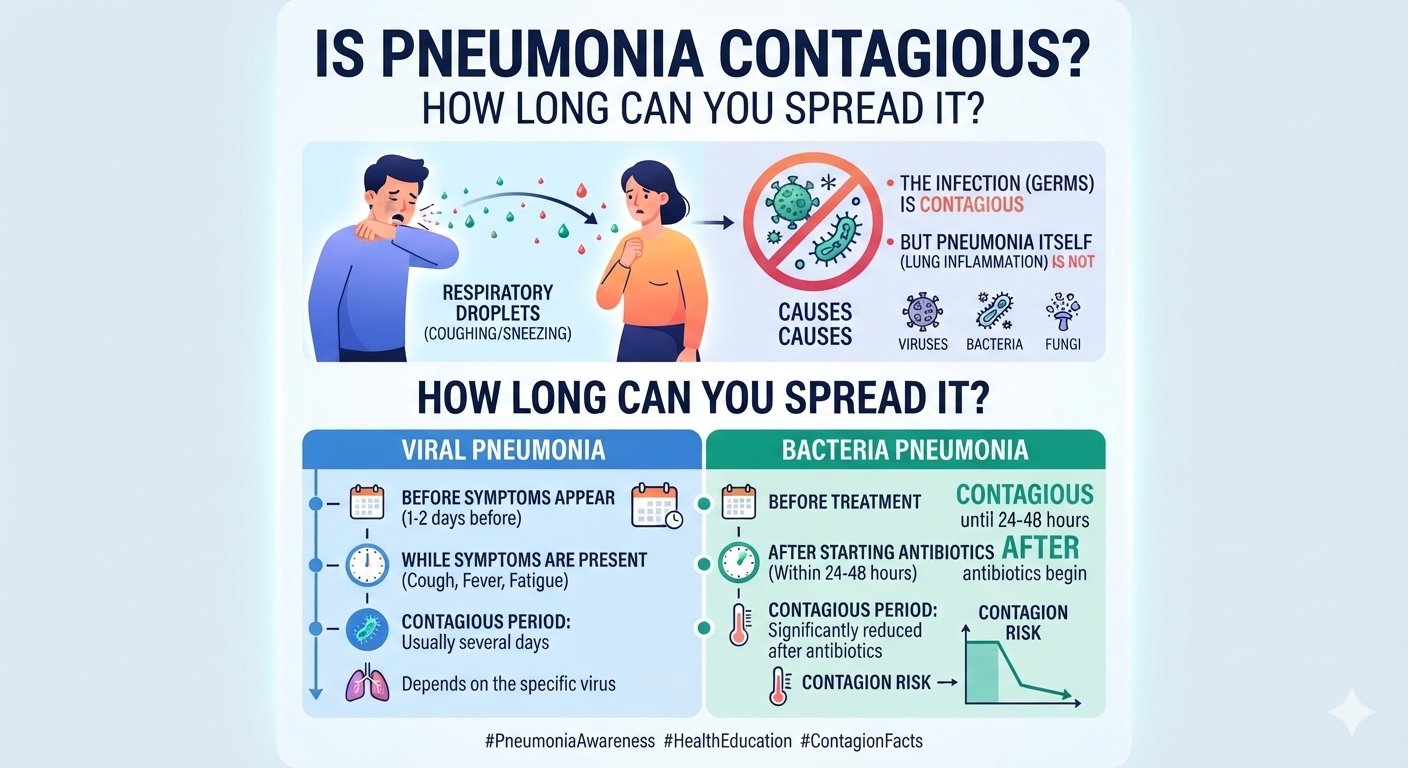
Stages 1–3 for bacterial; the entire symptomatic period for viral. See our full article on contagion.
Summary
Understanding pneumonia types and stages helps patients and families make sense of a confusing illness. The four main classifications are by setting (CAP, HAP, VAP), anatomy (lobar, broncho, interstitial), pathogen (bacterial, viral, fungal, aspiration), and pathological stage (congestion → red hepatization → grey hepatization → resolution). Severity is scored by CURB-65 or PSI in adults and WHO ladder in children. The right type and stage dictate the antibiotic, the care setting, and the recovery timeline.
Conclusion
When you leave your doctor’s clinic with a pneumonia diagnosis, ask three questions: What type is it? What stage? What is my severity score? The answers transform a frightening word into a plan. For patients and families in Nashik and surrounding areas, I offer in-clinic respiratory assessment and treatment at www.entspecialistinnashik.com.
📞 ENT Consultation & Respiratory Care
Dr. Sagar Rajkuwar (MS-ENT)
Prabha ENT Clinic, Ambad, Nashik
📱 7387590194 | 9892596635
🌐 www.entspecialistinnashik.com
References
- WHO — Pneumonia Fact Sheet
- CDC — Pneumonia
- NHS — Pneumonia
- ATS/IDSA — CAP Guidelines
- BTS — Pneumonia Guidelines
- NIH / NCBI — Community-Acquired Pneumonia
- Radiopaedia — Pneumonia
- Indian Academy of Pediatrics
Related Articles on Healthuseful
- Pneumonia Recovery Time at Home
- Can Pneumonia Be Cured Without Antibiotics?
- Pneumonia Symptoms in Toddlers
- Chest X-Ray Pneumonia Appearance
- Pneumonia Home Remedies Natural
- Is Pneumonia Contagious? How Long
- Early Signs of Pneumonia in Adults
- Walking Pneumonia Symptoms in Adults
- How Long Does Pneumonia Last?
- Pneumonia vs Bronchitis Symptoms
- Bacterial vs Viral Pneumonia
- Aspiration Pneumonia Symptoms in the Elderly
External Links
- YouTube: Healthuseful YouTube Channel
- Facebook: Positive Mind Healthcare Facebook
Medical Disclaimer: This article is for informational purposes only and does not substitute professional medical advice. Please consult a qualified healthcare provider for diagnosis and treatment.
Written by Dr. Sagar Rajkuwar, MS-ENT. Clinic: www.entspecialistinnashik.com | Nashik, Maharashtra, India | For appointments visit the clinic website.