By Dr Sagar Rajkuwar (MS-ENT) · Reviewed: 4 November 2026
Recognising deep vein thrombosis symptoms early is one of the most consequential skills any adult, carer or clinician can carry. A deep vein thrombosis (DVT) is a blood clot that forms inside a deep vein — most often in the calf or thigh, less commonly in the pelvis, abdomen or arms. Approximately 1 in 1,000 adults is affected every year; the risk rises with age. Dangerously, only around half of those with DVT have any obvious symptoms, which is why knowing the warning signs matters so much. Left untreated, a clot can break off and travel to the lungs, causing a pulmonary embolism (PE) — a life-threatening emergency.
This complete 2026 doctor’s guide walks through deep vein thrombosis symptoms as defined by the NHS, NICE NG158, Thrombosis UK and peer-reviewed vascular medicine. I cover the classical leg signs, less-common presentations (upper-extremity DVT, pelvic vein thrombosis, bilateral DVT), red flags that mean “call 999 now”, when to ask for an urgent GP appointment or NHS 111, how doctors confirm the diagnosis (Wells score, D-dimer, ultrasound), treatment overview and clear practical advice.
Table of Contents
- 1. What Is a Deep Vein Thrombosis?
- 2. Classical Leg Symptoms
- 3. Full List of Deep Vein Thrombosis Symptoms
- 4. Upper-Extremity & Pelvic DVT
- 5. Pulmonary Embolism — the Life-Threatening Red Flag
- 6. When to Call 999, NHS 111 or Your GP
- 7. How DVT Is Diagnosed (NICE NG158)
- 8. How DVT Is Treated
- 9. Complications & Long-Term Outlook
- 10. Prevention of DVT
- 11. Frequently Asked Questions
- 12. Summary & Conclusion
- 13. References
1. What Is a Deep Vein Thrombosis?
A deep vein thrombosis is a blood clot (thrombus) that forms inside one of the deep veins of the body. The deep veins run through the muscles — unlike the superficial veins visible just under the skin — and carry the majority of blood returning to the heart. The most common site is the calf (soleal or gastrocnemius veins), but DVT can extend upward into the popliteal vein behind the knee, the femoral vein in the thigh, the iliac veins in the pelvis, and the inferior vena cava in the abdomen.
Understanding the anatomy helps make sense of deep vein thrombosis symptoms. A clot blocks venous blood return, so blood pools behind it. This raises the pressure in the vein, pushes fluid out of the capillaries into surrounding tissue (oedema), stretches pain receptors in the vein wall, and causes the local skin to look red, warm and sometimes shiny. Approximately 90% of symptomatic DVTs occur in the legs; around 10% occur in the upper limbs (brachial, axillary or subclavian veins), often related to central venous catheters or thoracic outlet syndrome. Here is a short NHS explainer video on deep vein thrombosis symptoms, diagnosis and treatment:
2. Classical Leg Symptoms
The NHS summarises the classical deep vein thrombosis symptoms in the leg as:
- Throbbing or cramping pain in one leg (rarely both legs) — usually in the calf or thigh
- Swelling of one leg (rarely both legs)
- Warm skin around the painful area
- Red or darkened skin around the painful area — may appear dark red, purple, brown or blue depending on skin tone
- Swollen, hard, palpable veins that feel tender to touch
These five features are the clinical hallmarks of deep vein thrombosis symptoms but — critically — up to 50% of DVTs produce none of them. The clot may be present and silently extending while the leg looks relatively normal. This is why risk assessment matters just as much as symptom checking (see sections 7 and 10).
3. Full List of Deep Vein Thrombosis Symptoms
Beyond the classical five, the complete clinical spectrum of deep vein thrombosis symptoms as defined by NICE NG158, Thrombosis UK and peer-reviewed vascular medicine includes:
- Tenderness along the course of the deep vein on palpation
- Homan’s sign — pain in the calf on forced dorsiflexion of the foot (now considered unreliable; not routinely used)
- Pitting oedema — leaves a dent when you press the shin with a thumb
- A sensation of “fullness” or heaviness in the affected leg
- Dilated superficial collateral veins on the surface of the leg (chronic cases)
- Difference in calf circumference > 3 cm between the two legs (NICE criterion)
- Phlegmasia cerulea dolens — a severe, rare presentation where massive clotting causes the whole leg to become blue, swollen and intensely painful; a limb-threatening surgical emergency
- Phlegmasia alba dolens — “milk leg” (white leg) — rare, white-leg variant, classically in pregnancy
Notably, isolated calf pain without swelling is one of the commonest under-diagnosed presentations of deep vein thrombosis symptoms — it is easily mistaken for a pulled muscle or cramp. If calf pain persists longer than 48 hours after any possible trigger (long journey, surgery, immobility), seek medical review.
4. Upper-Extremity & Pelvic DVT
Around 10% of DVTs occur in the arm — upper-extremity DVT (UEDVT) — typically in the brachial, axillary or subclavian veins. The classical presentation of UEDVT deep vein thrombosis symptoms is unilateral arm swelling, discolouration, a feeling of heaviness or aching, and sometimes prominent chest-wall collateral veins. UEDVT is commonly associated with central venous catheters, PICC lines, cardiac pacemakers, and thoracic outlet syndrome (Paget-Schroetter syndrome in young, otherwise healthy people with repetitive overhead activity).
Pelvic vein thrombosis — clotting in the iliac veins — can present atypically, sometimes with lower abdominal pain as the only symptom. It is more common in pregnancy (especially left-sided due to anatomical compression by the right iliac artery, May-Thurner syndrome), in the post-partum period and after pelvic or abdominal surgery. If you have persistent one-sided pelvic or groin pain with any risk factor, seek assessment — pelvic DVT is often missed on calf-focused examination.
5. Pulmonary Embolism — the Life-Threatening Red Flag
The most dangerous complication of the full set of deep vein thrombosis symptoms is pulmonary embolism (PE) — when a piece of clot breaks off, travels through the heart and lodges in a pulmonary artery, obstructing blood flow to the lungs. PE is a medical emergency. The NHS urges immediate dialling of 999 if someone with possible DVT develops any of:
- Shortness of breath (new or sudden onset)
- Chest pain — may be sharp, worse on breathing in (pleuritic)
- Coughing up blood (haemoptysis)
- Fast heart rate (tachycardia, >100 beats per minute)
- Dizziness, fainting or collapse
- Clammy, pale or blue-tinged skin
Any combination of these — especially with known deep vein thrombosis symptoms in the leg — must prompt immediate 999 call. Do not drive to A&E yourself; ask someone to drive you or call an ambulance.
6. When to Call 999, NHS 111 or Your GP
The NHS pathway for deep vein thrombosis symptoms is straightforward:
- Call 999 immediately if you have any PE features — breathlessness, chest pain, coughing blood, collapse
- Request an urgent GP appointment or contact NHS 111 if you have suspected DVT symptoms (one-sided leg pain, swelling, redness, warmth, hard tender veins)
- If a doctor suspects DVT, the NHS pathway targets referral for ultrasound within 24 hours
- Do not massage a painful swollen leg — this may dislodge a clot
- Do not wait to see if symptoms resolve — early treatment prevents PE
7. How DVT Is Diagnosed (NICE NG158)
The modern NHS and NICE NG158 (2020, updated 2023) diagnostic algorithm for suspected deep vein thrombosis symptoms uses three steps:
7.1 Clinical Assessment & Wells Score
The two-level Wells score stratifies risk. Points are assigned for active cancer (+1), paralysis or recent plaster immobilisation (+1), bedridden > 3 days or major surgery within 12 weeks (+1), localised tenderness along deep veins (+1), entire leg swollen (+1), calf swelling > 3 cm vs the other leg (+1), pitting oedema confined to the symptomatic leg (+1), collateral superficial veins (+1), previous DVT (+1), and −2 if an alternative diagnosis is at least as likely as DVT.
- DVT likely: Wells score ≥ 2
- DVT unlikely: Wells score ≤ 1
7.2 D-Dimer Blood Test
If DVT is unlikely on Wells (≤1), a D-dimer is performed. A negative D-dimer effectively rules out DVT. A positive D-dimer prompts an ultrasound. D-dimer alone cannot confirm DVT because it rises in many conditions (infection, trauma, cancer, pregnancy, surgery, old age).
7.3 Doppler Ultrasound
The gold-standard confirmatory investigation for deep vein thrombosis symptoms is proximal leg vein ultrasound with compression Doppler. If veins are not compressible under the probe, a clot is present. If positive, anticoagulation starts immediately; if negative with a positive D-dimer and Wells ≥ 2, the ultrasound is repeated after 6-8 days. CT or MR venography is reserved for pelvic or vena-caval clots.
8. How DVT Is Treated
Once deep vein thrombosis symptoms are confirmed, the mainstay of treatment is anticoagulation. NICE NG158 recommends direct oral anticoagulants (DOACs) as first-line for most patients — apixaban or rivaroxaban, both started at an initial higher dose then stepped down. Edoxaban and dabigatran are alternatives (each after initial heparin cover). Warfarin remains the drug of choice in antiphospholipid syndrome. Low-molecular-weight heparin (LMWH) such as tinzaparin, enoxaparin or dalteparin is preferred in pregnancy and in some cancer-associated DVT.
Standard duration is a minimum of 3 months. Longer or indefinite anticoagulation is considered for recurrent or unprovoked DVT, active cancer, and thrombophilia. Compression stockings are no longer routinely recommended for acute DVT by NICE but may help post-thrombotic syndrome. Delay flights or long journeys for at least 2 weeks after starting anticoagulation. For life-threatening iliofemoral DVT or phlegmasia cerulea dolens, catheter-directed thrombolysis or thrombectomy may be considered.
9. Complications & Long-Term Outlook
Even after successful treatment of deep vein thrombosis symptoms, two important long-term complications can occur. Post-thrombotic syndrome (PTS) affects 20-50% of patients within 2 years — a chronic condition of leg swelling, pain, heaviness, skin changes and sometimes venous ulceration caused by persistent valve damage. Early effective anticoagulation and graduated compression stockings reduce the risk. The second complication is recurrent VTE — the risk is around 30% at 10 years, higher in unprovoked clots.
10. Prevention of DVT
Prevention of recurrent deep vein thrombosis symptoms or first-episode DVT is based on a combination of lifestyle and pharmacological measures. During hospital admission or after surgery, the NHS routinely assesses every adult patient for VTE risk and prescribes mechanical (compression stockings, intermittent pneumatic compression) and/or pharmacological prophylaxis (LMWH or DOAC) as indicated. On long journeys (flights more than 4 hours, train or car travel more than 6 hours), move about every 1-2 hours, flex your calves regularly, stay well hydrated, avoid excess alcohol, and if at higher risk, wear graduated compression stockings.
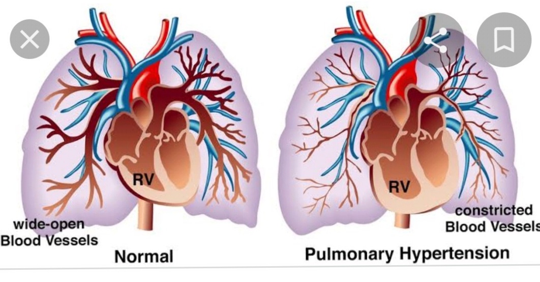
Stop smoking, maintain a healthy weight, manage diabetes and hypertension, and discuss alternatives to combined oral contraceptives or hormone replacement therapy (HRT) with your doctor if you have other clot risk factors. For related reading on this blog, see my articles on pulmonary hypertension complications, warning signs after childbirth, and surgical treatment of ischemic heart disease. See also my companion article on deep vein thrombosis causes.
11. Frequently Asked Questions (FAQs)
Q1. What are the first signs of a DVT in the leg?
The first deep vein thrombosis symptoms are usually throbbing or cramping pain in one leg (especially calf), one-sided swelling, warm skin, and red or darkened skin over the painful area. Not everyone has these; up to half of DVTs are silent.
Q2. Can DVT go away without treatment?
Small calf-vein clots can occasionally resolve spontaneously, but this is unpredictable and the risk of extension or pulmonary embolism (PE) is real. Any suspected DVT must be medically assessed; do not wait.
Q3. How quickly do DVT symptoms appear after a long flight?
Post-travel deep vein thrombosis symptoms usually appear within days to 4 weeks of the flight. Always be alert in the four weeks after any journey > 4 hours, particularly if you have other risk factors (older age, previous DVT, pregnancy, HRT, obesity, cancer, recent surgery).
Q4. Can DVT be painless?
Yes. Approximately 50% of DVTs are entirely asymptomatic or present with very mild discomfort only. This is why risk-factor awareness is important — see the companion article on deep vein thrombosis causes.
Q5. Is DVT common in both legs?
No. DVT is nearly always in one leg (unilateral). Bilateral leg swelling usually suggests a non-DVT cause (heart failure, kidney disease, venous insufficiency), but occasionally inferior vena cava thrombosis can produce bilateral symptoms.
Q6. Can you walk with a DVT?
Yes — contrary to older teaching, bed rest is no longer recommended once anticoagulation has started. Walking (within comfort) actually reduces leg swelling and improves recovery. Do not massage the leg.
Q7. What is a D-dimer test?
D-dimer is a blood test that measures a fibrin breakdown product. A negative D-dimer in a patient with low-probability Wells score effectively rules out DVT. A positive D-dimer is non-specific and prompts ultrasound confirmation.
12. Summary and Conclusion
Deep vein thrombosis symptoms are a critical skill for every adult to recognise. The classical signs — one-sided throbbing leg pain, swelling, warmth, red or darkened skin, hard tender veins — appear in only about half of cases, which is why risk-factor awareness and prompt medical evaluation of any suspicion are essential. The NHS pathway is clear: ask for an urgent GP appointment or call NHS 111 if symptoms suggest DVT; call 999 immediately for any features of pulmonary embolism (breathlessness, chest pain, coughing blood, collapse).
Modern NICE NG158 care involves a Wells-score-driven pathway with D-dimer blood testing and compression Doppler ultrasound. Treatment with direct oral anticoagulants (apixaban, rivaroxaban, edoxaban, dabigatran), low-molecular-weight heparin or warfarin as indicated prevents extension, embolism and most deaths. Long-term complications — post-thrombotic syndrome and recurrent VTE — can be minimised by early diagnosis, consistent anticoagulation and lifestyle modification. Prevention during hospital, after surgery and on long journeys is one of the quiet success stories of modern NHS care.
13. References
- NICE Guideline NG158. Venous thromboembolic diseases: diagnosis, management and thrombophilia testing. 2020, updated 2023.
- NHS. Deep vein thrombosis (DVT). nhs.uk/conditions/deep-vein-thrombosis-dvt (updated 2026).
- NHS Wales 111. DVT (deep vein thrombosis). 111.wales.nhs.uk, June 2026 update.
- Thrombosis UK. Venous thromboembolism patient information resources. 2026.
- Royal College of General Practitioners. Elearning module on VTE / NICE 2023 update. 2024-26.
- Wells P et al. Evaluation of D-dimer in the diagnosis of suspected deep vein thrombosis. NEJM 2003;349:1227-35.
- Khan F, Tritschler T, Kahn SR et al. Venous thromboembolism. The Lancet 2021;398(10294):64-77.
- UCLH. Deep vein thrombosis — easy-read patient information. 2026.
- NHS Resolution. Venous thromboembolism — did you know? 2023.
- Oxford Medical Education. Causes of deep vein thrombosis. 2024.
ENT Consultation & Surgery
Dr. Sagar Rajkuwar (MS-ENT)
Consultant ENT Specialist & Head-Neck Surgeon
Prabha ENT Clinic, Ambad, Nashik, Maharashtra, India
📱 7387590194 | 9892596635
🌐 www.entspecialistinnashik.com
Medical Disclaimer: This article is for educational purposes only and does not replace professional medical advice. Suspected deep vein thrombosis must be evaluated by a qualified clinician — ask for an urgent GP appointment or call NHS 111. In case of any symptom of pulmonary embolism (breathlessness, chest pain, coughing blood, collapse) call 999 immediately. Do not drive yourself to A&E.