
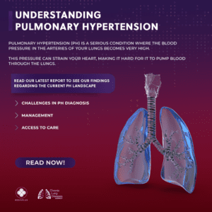
Pulmonary Hypertension Diagnosis-various aspects-
Pulmonary hypertension (PH) is identified through a series of tests, such as echocardiograms, right heart catheterizations, and pulmonary function assessments.
Tests
Echocardiogram: Employs sound waves to generate an image of the heart, which can reveal blood flow and pressure within the pulmonary arteries.
Right heart catheterization: A slender tube is inserted into a vein in the neck, arm, or groin and navigated into the pulmonary artery to assess blood pressure.
Pulmonary function tests: Evaluate the total air capacity of the lungs, how effectively they exchange oxygen, and the volume of air that moves in and out.
Chest X-ray: Produces images of the heart, lungs, and chest to identify additional lung issues.
Electrocardiogram (ECG or EKG): Monitors the heart’s electrical activity to identify any changes or damage.
Blood tests: Search for blood clots, anemia, or strain on the heart.
Cardiac MRI: Captures detailed images of the heart and blood vessels.
Symptoms
Signs of PH encompass:
Shortness of breath, particularly during physical activity.
Fatigue.
Swelling in the feet, legs, abdomen, or neck.
Dizziness or episodes of fainting.
Chest pain.
Heart palpitations.
Bluish tint in the lips and fingers.
Cough.
Hoarseness.
Diagnosis
Pulmonary hypertension is challenging to identify early because it is not frequently detected during a standard physical examination. Even when pulmonary hypertension has progressed, its symptoms resemble those of various other heart and lung diseases.
To identify pulmonary hypertension, a healthcare provider will examine you and inquire about your symptoms. You will probably be asked questions regarding your medical and family histories.
Tests
Tests conducted to assist in diagnosing pulmonary hypertension may comprise:
Blood tests. Blood tests can aid in determining the cause of pulmonary hypertension or indicate signs of complications.
Chest X-ray. A chest X-ray produces images of the heart, lungs, and chest. It might be employed to look for other lung conditions that could lead to pulmonary hypertension.
Electrocardiogram (ECG or EKG). This straightforward test documents the heart’s electrical activity. It can reveal alterations in the heartbeat.
Echocardiogram. Sound waves are utilized to generate moving images of the beating heart. An echocardiogram illustrates blood flow through the heart. This test may be performed to help diagnose pulmonary hypertension or to assess the effectiveness of treatments.
Occasionally, an echocardiogram is conducted while exercising on a stationary bike or treadmill to evaluate how activity impacts the heart. If you undergo this test, you might be requested to wear a mask that monitors how well the heart and lungs utilize oxygen and carbon dioxide.
Right heart catheterization. If an echocardiogram indicates pulmonary hypertension, this examination may be carried out to validate the diagnosis.
During this procedure, a cardiologist inserts a slender, flexible tube known as a catheter into a blood vessel, typically in the neck. The catheter is smoothly navigated into the lower right heart chamber and the pulmonary artery. A physician can then measure blood pressure in the main pulmonary arteries and the right ventricle.
Additional tests may be performed to evaluate the condition of the lungs and pulmonary arteries. The following tests may provide further information regarding the cause of pulmonary hypertension:
Exercise stress tests. These assessments frequently involve walking on a treadmill or biking on a stationary cycle while monitoring the heartbeat. They can illustrate how the heart responds to physical activity.
Computerized tomography (CT) scan. This test employs X-rays to create cross-sectional images of designated body areas. A dye known as contrast may be administered into a vein to enhance the visibility of blood vessels in the images.
A heart CT scan, referred to as a cardiac CT scan, can display the size of the heart and any blockages in the pulmonary arteries. It can assist in diagnosing lung disorders that may contribute to pulmonary hypertension, such as COPD or pulmonary fibrosis.
Magnetic resonance imaging (MRI). This examination utilizes magnetic fields and radio waves to create detailed images of the heart. It can depict blood flow within the pulmonary arteries and determine how well the right lower heart chamber is functioning.
Lung function assessment. For this assessment, you exhale into a specialized device. The device quantifies the volume of air the lungs can accommodate. It indicates how air circulates in and out of the lungs.
Sleep examination. A sleep examination evaluates brain activity, heart rate, blood pressure, oxygen saturation, and various other metrics while you are asleep. The assessment can assist in identifying sleep apnea, which may lead to pulmonary hypertension.
Ventilation/perfusion (V/Q) imaging. In this imaging, a radioactive tracer is administered through a vein (IV). The tracer illustrates blood circulation. You may also inhale a tracer that indicates airflow to the lungs. A V/Q imaging can reveal if blood clots are responsible for symptoms of pulmonary hypertension.
Lung tissue sampling. In rare cases, a tissue sample may be extracted from the lung to investigate a potential reason for pulmonary hypertension.
Genetic testing
Screening for genetic alterations that lead to pulmonary hypertension may be advised. If you possess these genetic alterations, other relatives may require screening as well.
Pulmonary hypertension functional classification
Upon confirming a diagnosis of pulmonary hypertension, the condition is categorized based on how the symptoms influence you and your capacity to perform daily activities.
Pulmonary hypertension may be classified into one of the following categories:
Class I. Pulmonary hypertension is identified, but there are no symptoms at rest or during exercise.
Class II. Symptoms are absent at rest. Routine tasks or activities like going to work or shopping can induce mild shortness of breath or minor chest discomfort. There’s a minor restriction in physical activities.
Class III. While at rest, it feels comfortable, but performing basic tasks such as bathing, dressing, or cooking leads to exhaustion, shortness of breath, and chest pain. The capacity for physical activity becomes significantly limited.
Class IV. Symptoms are present both at rest and during physical exertion. Any form of activity results in increasing discomfort.
Your medical team might employ a risk calculator that takes into account your symptoms and test outcomes to determine the appropriate type of treatment needed. This process is referred to as pulmonary hypertension risk stratification.
Care at Mayo Clinic
Our compassionate team of Mayo Clinic specialists is available to assist you with your health issues related to pulmonary hypertension.


