Varicose Veins Stages: From Early Spider Veins to Severe Venous Ulcers (CEAP Classification)
Article author: Dr. Sagar Rajkuwar, MS-ENT
Introduction
Varicose veins are more than just enlarged or twisted blood vessels visible beneath the skin. They can represent an underlying problem with the valves that control blood flow through the veins of the legs. As this condition progresses, patients may develop symptoms such as leg heaviness, swelling, skin discolouration, eczema and, in advanced cases, painful venous ulcers. Understanding the varicose veins stages helps patients recognise the severity of their condition and seek appropriate medical attention before complications develop. In the early stages, the problem may appear as small spider veins or slightly enlarged superficial veins. As venous disease becomes more advanced, fluid may accumulate in the legs, skin changes may develop and wounds may form around the ankles. Doctors use an internationally recognised system called the Clinical-Etiological-
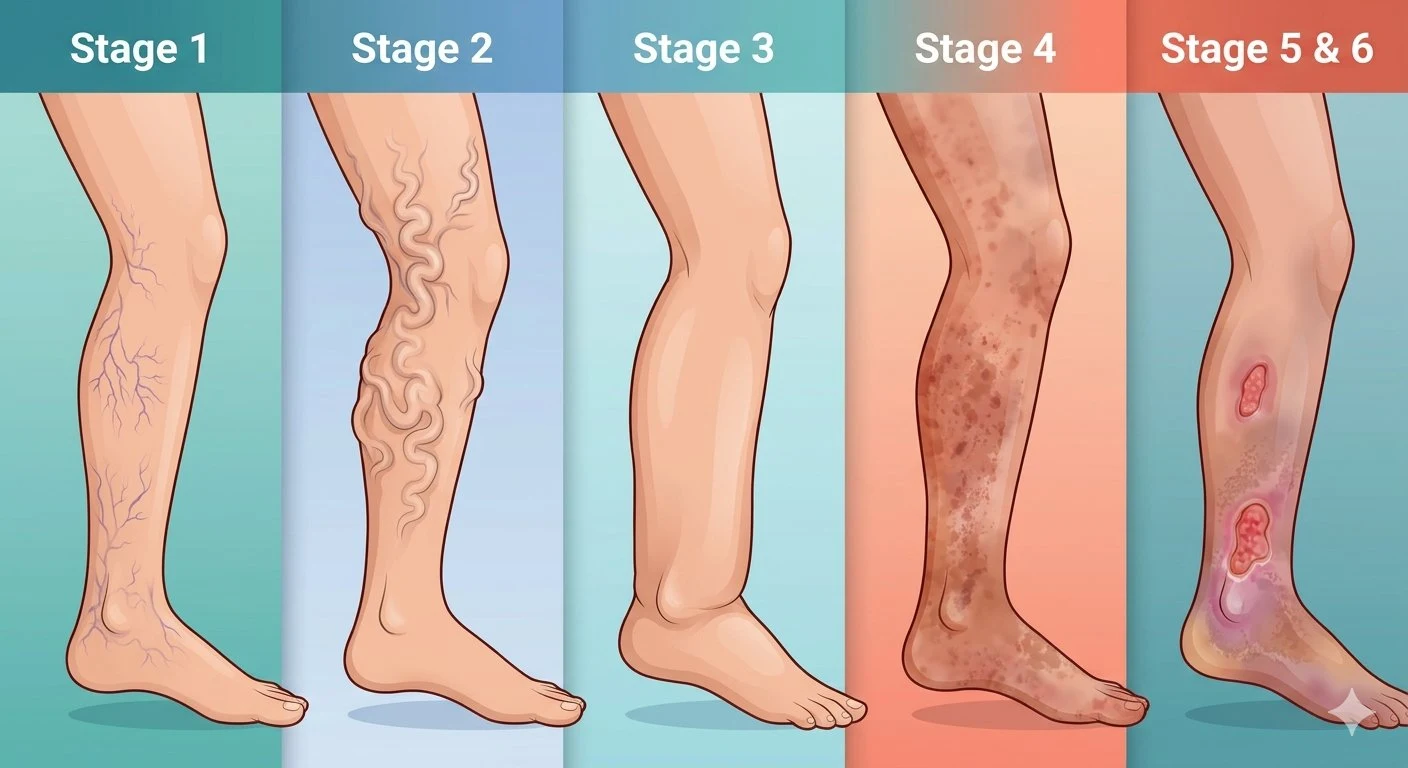
- C0: No visible or palpable signs of venous disease.
- C1: Spider veins and reticular veins.
- C2: Established varicose veins.
- C3: Leg swelling or oedema.
- C4: Skin and subcutaneous tissue changes.
- C5: Healed venous ulcer.
- C6: Active venous ulcer.
An important distinction is that these classes describe the clinical condition rather than a fixed sequence that every patient must follow. Some patients have visible varicose veins for many years without developing ulcers, while others develop significant swelling or skin changes. This comprehensive guide explains the different stages, their symptoms, causes, complications, diagnostic investigations and appropriate treatment options. It also explains when varicose veins require specialist assessment and which warning signs should never be ignored.
1. What Are Varicose Veins?
Varicose veins are enlarged, twisted and dilated superficial veins, most commonly affecting the legs. Normally, veins transport blood from the lower limbs towards the heart. Small structures called venous valves prevent blood from flowing backwards under the influence of gravity. When these valves become incompetent, blood flows backwards and accumulates inside the veins. This condition is known as venous reflux. The increased pressure within the superficial veins causes them to enlarge, become twisted and sometimes protrude beneath the skin.
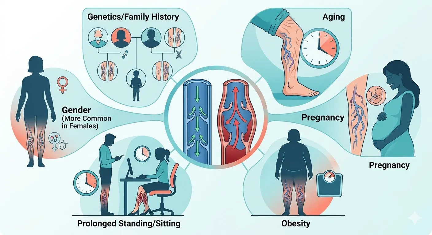
Why do varicose veins develop?
Several factors contribute to the development of venous disease:
- Family history.
- Increasing age.
- Pregnancy.
- Obesity.
- Prolonged standing.
- Previous deep vein thrombosis.
- Weakness of venous walls and valves.
- Reduced calf-muscle activity.
What is chronic venous disease?
Chronic venous disease (CVD) is a broad term describing abnormalities of the veins that interfere with normal venous circulation. It includes conditions ranging from small spider veins to advanced chronic venous insufficiency with skin damage and leg ulcers. Chronic venous insufficiency (CVI) generally refers to more advanced venous disease, particularly when venous hypertension produces swelling, skin changes or ulceration. Understanding this distinction is important because not every person with visible varicose veins has advanced venous insufficiency.
2. What Are the Different Stages of Varicose Veins?
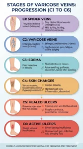
The internationally recognised CEAP classification provides a standardised way of describing chronic venous disease. Although patients often search for mild, moderate and severe varicose veins, the CEAP system offers a more detailed clinical assessment. CEAP clinical classification at a glance
| Class | Clinical description | Main finding |
|---|---|---|
| C0 | No visible or palpable venous signs | Symptoms may still be present |
| C1 | Spider or reticular veins | Small superficial vessels |
| C2 | Varicose veins | Enlarged, twisted veins |
| C3 | Oedema | Leg or ankle swelling |
| C4 | Skin and tissue changes | Pigmentation, eczema or fibrosis |
| C5 | Healed venous ulcer | Previously ulcerated skin |
| C6 | Active venous ulcer | Open venous wound |
The following sections explain each clinical class in detail.
3. Stage C0: No Visible Signs of Venous Disease
C0 is the earliest clinical category in the CEAP classification. At this stage, there are no visible or palpable signs of venous disease. However, some patients may experience symptoms associated with venous dysfunction.
Common symptoms
- Leg heaviness.
- Aching.
- Discomfort after prolonged standing.
- A sensation of swelling.
- Fatigue in the legs.
- Occasional itching or burning.
The symptoms may become more noticeable towards the evening.
Why can symptoms occur without visible varicose veins?
Venous dysfunction may sometimes exist before obvious surface changes become apparent. However, these symptoms are not specific to venous disease. Similar complaints can occur due to:
- Musculoskeletal disorders.
- Peripheral neuropathy.
- Joint problems.
- Peripheral arterial disease.
- Medication-related swelling.
- Other causes of leg oedema.
Therefore, symptoms alone cannot establish a diagnosis of chronic venous disease.
What does C0s mean?
In the CEAP classification, the letter S indicates that symptoms are present. C0s therefore means that a patient has symptoms attributed to venous disease but no visible or palpable clinical signs. The letter A indicates an asymptomatic presentation.
What should patients do?
Persistent or unexplained symptoms should be assessed by a healthcare professional. If the clinical history suggests venous disease, a venous duplex ultrasound may be recommended.
4. Stage C1: Spider Veins and Reticular Veins
EARLY VISIBLE VENOUS CHANGES C1 is characterised by small superficial blood vessels visible beneath the skin. There are two important types:
- Telangiectasias (spider veins).
- Reticular veins.
4.1 Spider veins
Spider veins are small, dilated blood vessels measuring less than 1 mm in diameter. They may appear as:
- Red lines.
- Blue lines.
- Purple web-like patterns.
- Small branching clusters.
They commonly develop around the thighs, knees and lower legs.
4.2 Reticular veins
Reticular veins are slightly larger superficial veins, generally measuring between 1 mm and less than 3 mm in diameter. They may appear blue or green beneath the skin. Unlike large varicose veins, they usually do not protrude significantly.
Do spider veins indicate serious venous disease?
Not necessarily. Many people have spider veins without significant underlying chronic venous insufficiency. However, some patients have associated superficial venous reflux. A clinical assessment helps determine whether further investigation is necessary.
Symptoms of C1 disease
Many patients have no symptoms. Others may experience:
- Mild burning.
- Itching.
- Local discomfort.
- Cosmetic concerns.
Treatment options
Treatment depends on the patient’s symptoms and preferences. Options may include:
- Sclerotherapy.
- Transcutaneous laser treatment.
- Appropriate management of associated venous reflux when present.
Not every patient requires medical intervention.
5. Stage C2: Established Varicose Veins
VISIBLE ENLARGED AND TWISTED VEINS C2 represents established varicose veins. At this stage, superficial veins become enlarged, dilated and tortuous. They may protrude beneath the skin and become more noticeable when the patient stands.
Appearance of C2 varicose veins
Typical findings include:
- Bulging blue or purple veins.
- Twisted superficial blood vessels.
- Rope-like veins along the calf or thigh.
- Multiple enlarged venous branches.
- Prominent veins around the knees.
Symptoms of C2 disease
Patients may experience:
- Leg heaviness.
- Aching.
- Pain after prolonged standing.
- Itching.
- Burning sensations.
- Night-time cramps.
- Mild ankle swelling in some cases.
Symptoms may improve after walking, resting or elevating the legs.
Why do veins become enlarged?
The main mechanism is venous valve incompetence. When valves fail to close properly, blood flows backwards. This increases pressure inside the superficial venous system. Over time, the affected veins become progressively dilated.
Which veins are commonly affected?
The important superficial venous trunks include:
- Great saphenous vein (GSV).
- Small saphenous vein (SSV).
- Anterior accessory great saphenous vein (AAGSV).
Incompetent tributary and perforator veins may also contribute.
Can C2 varicose veins be treated?
Yes. Treatment options include:
- Endovenous laser ablation.
- Radiofrequency ablation.
- Ultrasound-guided foam sclerotherapy.
- Cyanoacrylate glue closure.
- Mechanochemical ablation.
- Ambulatory phlebectomy.
The choice depends on the venous anatomy and ultrasound findings.
What is recurrent varicose vein disease?
The updated CEAP classification includes C2r, which describes recurrent varicose veins. These are varicose veins that develop again after previous treatment. Recurrence may occur because of:
- New venous reflux.
- Recanalisation of a previously treated vein.
- Development of reflux in another superficial vein.
- Progression of the underlying disease.
6. Stage C3: Leg Swelling or Oedema
SWELLING DUE TO FLUID ACCUMULATION C3 is characterised by oedema, meaning abnormal accumulation of fluid within the tissues. In chronic venous disease, increased pressure within the veins can cause fluid to move from the blood vessels into surrounding tissues. The swelling is commonly seen around the ankles and lower legs.
Symptoms of C3 disease
Patients may notice:
- Swelling around one or both ankles.
- Increased leg circumference.
- Tightness around the lower leg.
- Heaviness.
- Discomfort after standing.
- Shoes becoming tighter towards the evening.
Swelling may improve after overnight rest or leg elevation.
Why does venous oedema develop?
Under normal circumstances, the venous and lymphatic systems work together to maintain fluid balance. When venous pressure increases, more fluid moves into the surrounding tissues. If the lymphatic system cannot remove this additional fluid efficiently, oedema develops.
Is all leg swelling caused by varicose veins?
No. This is an important clinical distinction. Other possible causes include:
- Deep vein thrombosis.
- Heart failure.
- Kidney disease.
- Liver disease.
- Lymphoedema.
- Medication-related oedema.
- Infection.
- Prolonged immobility.
Sudden swelling affecting one leg, particularly with pain, warmth or tenderness, requires prompt medical evaluation to exclude DVT.
Treatment of C3 disease
Treatment depends on the underlying cause. For venous oedema, options may include:
- Treating significant superficial venous reflux.
- Graduated compression when appropriate.
- Regular walking.
- Calf-muscle exercises.
- Leg elevation.
- Weight management.
Compression should be prescribed after considering arterial circulation, particularly in patients with diabetes or peripheral arterial disease.
7. Stage C4: Skin Changes Due to Chronic Venous Disease
ADVANCED CHRONIC VENOUS INSUFFICIENCY C4 represents an important stage of chronic venous disease. At this stage, prolonged venous hypertension has affected the skin and underlying tissues. The changes may become permanent if the underlying venous problem remains untreated. The updated CEAP classification divides C4 into three subcategories.
7.1 C4a: Pigmentation and Venous Eczema
The main features are:
- Brownish skin discolouration.
- Venous eczema.
- Dryness.
- Scaling.
- Itching.
- Redness and inflammation.
Why does skin pigmentation develop?
Increased venous pressure can cause small blood vessels to leak red blood cells into surrounding tissues. When these cells break down, haemosiderin, an iron-containing pigment, accumulates in the skin. This produces characteristic brown or rusty-coloured pigmentation. The changes are often most prominent around the ankles.
What is venous eczema?
Venous eczema, also called stasis dermatitis, is an inflammatory skin condition associated with chronic venous hypertension. Symptoms include:
- Itchy skin.
- Redness.
- Scaling.
- Dryness.
- Irritation.
- Sometimes fluid leakage from inflamed skin.
Patients should seek medical advice because venous eczema can resemble other dermatological conditions.
7.2 C4b: Lipodermatosclerosis and Atrophie Blanche
This category describes more advanced changes involving the skin and subcutaneous tissues.
What is lipodermatosclerosis?
Lipodermatosclerosis is a chronic inflammatory and fibrotic condition affecting the lower leg. The skin and underlying tissues may become:
- Thickened.
- Hard.
- Painful.
- Tightly adherent.
- Discoloured.
The lower leg may develop a characteristic inverted champagne-bottle appearance, with narrowing above the ankle and relative enlargement of the calf.
What is atrophie blanche?
Atrophie blanche describes small, pale or whitish scar-like areas surrounded by discoloured skin and sometimes small dilated blood vessels. It is associated with chronic venous disease and can indicate significant underlying tissue damage.
7.3 C4c: Corona Phlebectatica
Corona phlebectatica is a fan-shaped arrangement of small dilated veins around the ankle and foot. It may appear as blue or red superficial vessels. This is an important sign of advanced chronic venous disease. It is specifically recognised in the updated CEAP classification.
Why is C4 clinically important?
Skin changes indicate that venous hypertension is affecting tissue health. Patients should not regard persistent pigmentation, eczema or skin hardening as merely cosmetic problems. Early vascular assessment can help identify treatable reflux and reduce the risk of further complications.
8. Stage C5: Healed Venous Leg Ulcer
PREVIOUS ULCER HAS HEALED C5 indicates that a patient previously had a venous leg ulcer, but the ulcer has now healed. Although the wound is closed, the underlying venous disease may remain.
What is a venous leg ulcer?
A venous leg ulcer is a chronic wound caused by prolonged venous hypertension and impaired tissue healing. It commonly develops in the lower leg, particularly around the medial ankle.
What does a healed ulcer look like?
The affected area may show:
- Scar tissue.
- Brownish pigmentation.
- Thin or fragile skin.
- Residual eczema.
- Local thickening.
- Persistent swelling.
Why can ulcers recur after healing?
The wound may heal while the underlying venous reflux remains untreated. Other factors contributing to recurrence include:
- Persistent venous hypertension.
- Poor mobility.
- Inadequate compression where indicated.
- Obesity.
- Previous DVT.
- Associated arterial disease.
- Poor wound-care practices.
How can recurrence be reduced?
Important measures include:
- Appropriate management of underlying venous reflux.
- Compression therapy when arterial circulation permits.
- Regular walking.
- Maintaining healthy skin.
- Avoiding prolonged immobility.
- Regular clinical follow-up.
- Managing associated medical conditions.
Patients with a healed venous ulcer should continue medical follow-up because recurrence is possible.
9. Stage C6: Active Venous Leg Ulcer
MOST ADVANCED CLINICAL CLASS C6 represents an active venous leg ulcer. It is the most advanced clinical category in the CEAP classification. At this stage, chronic venous hypertension has caused significant damage to the skin and surrounding tissues, resulting in an open wound.
Where do venous ulcers usually develop?
They commonly develop around the medial lower leg, particularly between the ankle and knee. The area near the medial malleolus is a frequent location.
Symptoms of C6 disease
Patients may experience:
- An open wound.
- Persistent discharge.
- Pain or tenderness.
- Swelling.
- Brownish skin pigmentation.
- Thickened surrounding skin.
- Venous eczema.
- Delayed wound healing.
The ulcer may have irregular edges and may produce fluid.
Why do venous ulcers develop?
Prolonged venous hypertension interferes with normal microcirculation and tissue function. Inflammatory changes, oedema and alterations in the skin environment impair wound healing. The result is a chronic wound that may remain open for weeks or months.
Can venous ulcers become infected?
Yes. Signs of infection may include:
- Increasing pain.
- Increasing redness or warmth.
- Pus or unpleasant discharge.
- Fever.
- Increasing swelling.
- Rapid deterioration of the wound.
A suspected infected venous ulcer requires prompt medical assessment.
How are active venous ulcers treated?
Management usually involves several approaches.
A. Wound care
The wound is assessed, cleaned and dressed appropriately. Dressings are selected according to the amount of discharge, wound condition and surrounding skin.
B. Compression therapy
Compression is an important part of venous ulcer treatment when significant arterial insufficiency has been excluded. It helps reduce venous hypertension and oedema. However, compression must be selected according to arterial circulation and the patient’s clinical condition.
C. Treatment of underlying venous reflux
Patients may benefit from treatment of superficial venous reflux. Procedures may include:
- Endovenous laser ablation.
- Radiofrequency ablation.
- Foam sclerotherapy.
- Other appropriate venous interventions.
D. Management of associated conditions
The doctor may also address:
- Diabetes.
- Nutritional deficiencies.
- Obesity.
- Infection.
- Reduced mobility.
- Arterial insufficiency.
Why is early treatment important?
An active venous ulcer can significantly affect mobility, sleep and quality of life. Prompt specialist management may improve healing and reduce the risk of recurrence.
10. Is There a Stage C7 in Varicose Veins?
No. The standard clinical component of the CEAP classification ranges from C0 to C6. C6 represents an active venous ulcer. There is no standard C7 clinical class in the recognised CEAP system. Patients may sometimes encounter descriptions such as mild, moderate, severe or end-stage varicose veins. These terms are useful for general communication but are not substitutes for formal CEAP classification.
11. Do Varicose Veins Always Progress From C1 to C6?
No. This is one of the most important points patients should understand. The CEAP classification describes the clinical condition at the time of examination. It does not mean that every person with spider veins will eventually develop an active venous ulcer. For example:
- A patient may remain in C1 for many years.
- Another patient may have symptomatic C2 disease without oedema.
- Some patients develop C3 swelling.
- Others may develop C4 skin changes.
- A smaller group develops venous ulcers.
Disease progression depends on multiple factors, including venous anatomy, underlying reflux, previous thrombosis, age, mobility, obesity and associated medical conditions.
Can someone have C4 disease without severe pain?
Yes. The severity of visible signs and the intensity of symptoms do not always correspond. Some patients with advanced skin changes experience relatively little pain, while others with less visible disease may have considerable discomfort. Therefore, clinical examination is more informative than relying on pain alone.
12. What Are the Different Types of CEAP Classification?
The full CEAP system contains four components. It is not limited to the clinical stage.
12.1 Clinical classification (C)
This describes the visible or palpable findings. It ranges from C0 to C6.
12.2 Etiological classification (E)
This describes the underlying cause. Important categories include:
| Category | Meaning |
|---|---|
| Ec | Congenital |
| Ep | Primary venous disease |
| Es | Secondary venous disease |
| En | No venous cause identified |
Primary venous disease develops due to abnormalities such as primary valve incompetence. Secondary venous disease may develop after conditions such as previous DVT or trauma.
12.3 Anatomical classification (A)
This describes the location of the affected veins. The main categories are:
- As: Superficial veins.
- Ad: Deep veins.
- Ap: Perforator veins.
- An: No venous location identified.
12.4 Pathophysiological classification (P)
This describes the underlying mechanism.
| Category | Meaning |
|---|---|
| Pr | Reflux |
| Po | Obstruction |
| Pr,o | Reflux and obstruction |
| Pn | No pathophysiology identified |
This additional information helps vascular specialists understand the underlying venous abnormality.
13. What Is the Difference Between Mild, Moderate and Severe Varicose Veins?
Patients frequently use these terms when describing their condition. Although they are not formal substitutes for CEAP classes, they can be used to explain the general clinical picture.
| General description | Typical findings | Approximate CEAP context |
|---|---|---|
| Mild | Spider veins and small reticular veins | C1 |
| Moderate | Visible enlarged varicose veins | C2 |
| More advanced | Leg or ankle swelling | C3 |
| Severe | Skin pigmentation, eczema or fibrosis | C4 |
| Advanced complicated disease | Healed or active venous ulcers | C5–C6 |
This is a simplified patient-education guide, not a validated staging system. A patient may have symptoms at any clinical class, and treatment decisions require individual assessment.
14. How Are Different Stages of Varicose Veins Diagnosed?
Diagnosis involves a clinical examination and, when indicated, venous imaging.
14.1 Medical history
The doctor asks about:
- Duration of symptoms.
- Family history.
- Previous DVT.
- Pregnancy history.
- Previous vein procedures.
- Occupation.
- Prolonged standing.
- Leg swelling.
- Previous ulcers.
- Associated medical conditions.
14.2 Physical examination
The legs are examined for:
- Spider veins.
- Reticular veins.
- Enlarged superficial veins.
- Oedema.
- Pigmentation.
- Venous eczema.
- Skin hardening.
- Healed or active ulcers.
14.3 Venous duplex ultrasound
Duplex ultrasound is the most important investigation for assessing superficial venous reflux and planning treatment. It helps identify:
- Great saphenous vein incompetence.
- Small saphenous vein incompetence.
- Incompetent tributaries.
- Perforator reflux.
- Deep vein obstruction.
- Evidence of previous or current thrombosis.
The examination should assess venous flow and valve function, often with the patient standing or in an appropriately dependent position.
14.4 Ankle-brachial pressure index
Before applying significant compression therapy, particularly in patients with ulcers or suspected arterial disease, doctors may assess arterial circulation. The ankle-brachial pressure index (ABPI) compares blood pressure measured at the ankle with blood pressure measured at the arm. A reduced value may indicate peripheral arterial disease. This is important because compression can be harmful in certain patients with severe arterial insufficiency.
15. When Should You Consult a Vascular Specialist?
You should seek medical assessment if you experience:
- Persistent leg heaviness.
- Pain associated with visible varicose veins.
- Increasing swelling.
- Skin pigmentation around the ankles.
- Venous eczema.
- Hard and painful superficial veins.
- A healed venous ulcer.
- A wound that has not healed within two weeks.
- Recurrent bleeding from varicose veins.
According to NICE guidance, symptomatic varicose veins, skin changes associated with chronic venous insufficiency, superficial vein thrombosis with suspected venous incompetence, and healed or active venous ulcers are reasons for referral to a vascular service.
When is emergency medical care necessary?
Seek emergency assistance for: Uncontrolled bleeding: Varicose veins close to the skin can bleed significantly after injury. Lie down, elevate the leg and apply firm direct pressure while obtaining urgent medical help. Possible DVT: Sudden one-sided leg swelling, pain, warmth or tenderness requires urgent assessment. Possible pulmonary embolism: Sudden breathlessness, chest pain, coughing up blood, fainting or collapse requires immediate emergency medical attention.
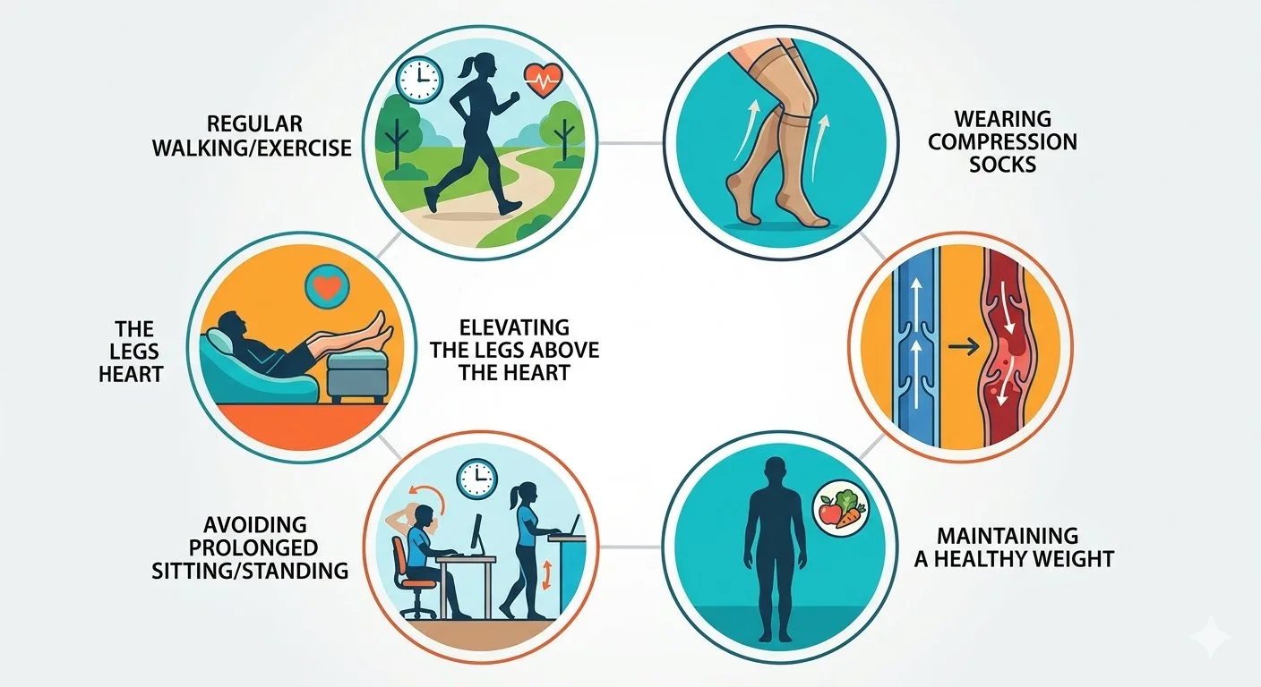
16. Can Varicose Veins Be Prevented From Progressing?
Not every case can be prevented, especially when genetic factors contribute. However, appropriate lifestyle measures may help reduce symptoms and support venous circulation.
16.1 Regular walking
Walking activates the calf-muscle pump, helping return blood towards the heart.
16.2 Avoid prolonged standing
People who stand for long periods should take regular movement breaks.
16.3 Avoid prolonged sitting
Regular walking and ankle movements help reduce venous stasis.
16.4 Maintain a healthy weight
Excess body weight can increase venous pressure.
16.5 Elevate the legs
Leg elevation while resting may help reduce swelling.
16.6 Compression stockings
Appropriately prescribed compression stockings may help selected patients manage swelling and discomfort. However, they do not permanently correct faulty venous valves.
16.7 Treat underlying venous reflux
Patients with symptomatic venous reflux may benefit from appropriate vascular intervention. The decision should be based on clinical examination and duplex ultrasound.
17. What Is the Best Treatment for Each Stage of Varicose Veins?
Treatment depends on the clinical class, symptoms, anatomy and presence of complications. The following table provides a simplified overview.
| CEAP class | Possible management |
|---|---|
| C0 | Investigate alternative causes of symptoms; assess venous disease if suspected |
| C1 | Reassurance, lifestyle measures and selected cosmetic treatments |
| C2 | Duplex ultrasound and consideration of endovenous treatment when symptomatic reflux is confirmed |
| C3 | Evaluate oedema causes, treat venous reflux and consider appropriate compression |
| C4 | Specialist vascular assessment, skin care and treatment of underlying venous hypertension |
| C5 | Ulcer recurrence prevention, compression where appropriate and venous intervention when indicated |
| C6 | Wound care, appropriate compression after arterial assessment and treatment of underlying venous disease |
Important treatment procedures
Depending on the ultrasound findings, vascular specialists may recommend:
- Endovenous laser ablation (EVLA).
- Radiofrequency ablation (RFA).
- Ultrasound-guided foam sclerotherapy.
- Cyanoacrylate glue closure.
- Mechanochemical ablation.
- Ambulatory phlebectomy.
- Conventional surgery in selected cases.
For patients with active venous ulcers and superficial venous reflux, early treatment of the underlying reflux alongside appropriate compression and wound care may improve ulcer healing. The EVRA trial provided important evidence supporting early endovenous treatment in appropriately selected patients with venous leg ulcers.
18. Can Varicose Veins Be Cured Permanently?
Treatment can successfully close or remove the abnormal veins responsible for symptoms. However, no procedure guarantees that new varicose veins will never develop. The underlying tendency towards venous valve dysfunction may persist. Recurrence can occur due to:
- New venous reflux.
- Recanalisation.
- Progression of chronic venous disease.
- Development of new incompetent veins.
Regular follow-up and appropriate management of associated risk factors are important.
19. Frequently Asked Questions About Varicose Veins Stages
1. What are the six stages of varicose veins?
The commonly described clinical stages are C1 spider or reticular veins, C2 established varicose veins, C3 oedema, C4 skin changes, C5 healed venous ulcer and C6 active venous ulcer. The full CEAP classification also includes C0, which indicates no visible or palpable signs.
2. What is the earliest stage of varicose veins?
C0 is the earliest clinical category, with no visible or palpable signs of venous disease. C1 is the earliest category with visible superficial venous changes, such as spider veins and reticular veins.
3. What does stage C1 mean?
C1 describes telangiectasias and reticular veins. These are small superficial blood vessels that may appear red, blue or purple beneath the skin.
4. What does stage C2 mean?
C2 describes established varicose veins. These are enlarged, dilated and twisted superficial veins, usually visible on the legs.
5. What does stage C3 mean?
C3 indicates oedema, or swelling, caused by fluid accumulation in the lower limbs. Chronic venous hypertension is one possible cause.
6. What does stage C4 mean?
C4 indicates skin and subcutaneous tissue changes associated with chronic venous disease. These may include pigmentation, venous eczema, lipodermatosclerosis, atrophie blanche and corona phlebectatica.
7. What is stage C5?
C5 means that a patient previously had a venous leg ulcer, but the wound has healed.
8. What is stage C6?
C6 is the most advanced clinical category in the CEAP system. It describes an active venous leg ulcer.
9. Are stage 1 varicose veins dangerous?
Small spider veins are generally not dangerous. However, patients with persistent symptoms or other signs of venous disease may require clinical assessment.
10. Can stage 2 varicose veins become severe?
Some patients develop progressive venous disease, while others remain stable for many years. Progression is not inevitable.
11. Can stage 3 varicose veins be treated?
Yes. Treatment may include management of venous reflux, appropriate compression, walking and leg elevation, depending on the cause of swelling.
12. Are stage 4 varicose veins serious?
C4 indicates skin changes caused by chronic venous disease. It deserves specialist assessment because persistent venous hypertension may increase the risk of ulceration.
13. Can stage 5 varicose veins become stage 6 again?
Yes. A previously healed venous ulcer may recur, resulting in an active ulcer classified as C6r when the ulcer is recurrent.
14. Can stage 6 varicose veins heal?
Yes. Many venous ulcers can heal with appropriate wound care, compression when safe, and management of underlying venous reflux. Healing may take several weeks or longer.
15. Is CEAP classification used worldwide?
Yes. CEAP is an internationally recognised classification system used by vascular specialists and researchers to describe chronic venous disease.
16. Does CEAP C6 mean the patient has DVT?
No. C6 describes an active venous leg ulcer. DVT is a separate condition involving thrombosis in the deep venous system, although previous DVT can contribute to chronic venous disease.
17. Can varicose veins cause permanent skin damage?
Yes. Advanced chronic venous hypertension may cause pigmentation, fibrosis, skin thickening and ulceration.
18. Does every patient with C2 varicose veins need laser treatment?
No. Treatment depends on symptoms, venous reflux, clinical findings and patient preference. Some patients may be managed conservatively, while others benefit from intervention.
19. Can compression stockings prevent progression to C6?
Compression may help control venous symptoms and oedema in selected patients, but it does not guarantee prevention of disease progression. Appropriate treatment of underlying venous reflux may also be necessary.
20. Which test determines the stage of varicose veins?
Clinical examination determines the clinical CEAP category. Duplex ultrasound helps identify venous reflux, obstruction and the affected veins. Both provide important information.
21. What is the difference between C4a and C4b?
C4a includes pigmentation and eczema. C4b includes lipodermatosclerosis and atrophie blanche. C4c is the additional category for corona phlebectatica.
22. Is there a C7 stage of varicose veins?
No. The standard CEAP clinical classification ranges from C0 to C6.
23. Can varicose veins occur without pain?
Yes. Many patients have visible varicose veins without significant discomfort. Others experience pain, heaviness, itching or swelling.
24. When should a patient with varicose veins visit a doctor?
Consult a doctor for persistent pain, swelling, skin changes, hard painful veins, bleeding or a non-healing wound. Sudden one-sided swelling or symptoms of pulmonary embolism require urgent or emergency assessment.
20. Summary: Varicose Veins Stages
The varicose veins stages are clinically described using the CEAP classification. The seven clinical categories are:
- C0: No visible or palpable venous signs.
- C1: Spider veins and reticular veins.
- C2: Established varicose veins.
- C3: Leg or ankle oedema.
- C4: Skin and tissue changes, including pigmentation, eczema and fibrosis.
- C5: Healed venous ulcer.
- C6: Active venous ulcer.
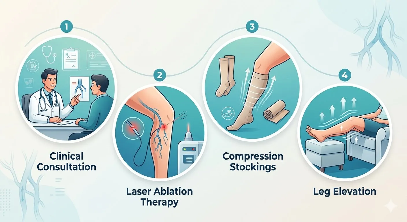
The most important point is that these categories describe clinical findings rather than a guaranteed sequence of progression. Some patients remain in the early stages for years, whereas others develop chronic venous insufficiency and complications. Duplex ultrasound is important for identifying venous reflux and planning treatment. Lifestyle changes, walking, leg elevation and appropriate compression may help manage symptoms. Patients with symptomatic venous reflux may benefit from endovenous laser treatment, radiofrequency ablation, foam sclerotherapy or other specialist procedures. Advanced skin changes, bleeding and venous ulcers require timely medical assessment.
21. Conclusion
Varicose veins can range from small superficial spider veins to advanced chronic venous disease involving skin damage and active venous ulcers. Understanding the CEAP classification helps patients and healthcare professionals describe the clinical severity of the condition and plan appropriate investigations and treatment. Although early varicose veins may cause mainly cosmetic concerns, persistent symptoms, swelling, pigmentation and eczema should not be ignored. The presence of a healed or active venous ulcer is particularly important because it indicates advanced chronic venous disease. Early specialist assessment, appropriate duplex ultrasound and individualised treatment can help relieve symptoms, manage complications and improve quality of life. Patients should remember that varicose veins do not necessarily progress through every stage. The most appropriate treatment depends on the underlying venous abnormality, symptoms and individual clinical circumstances.
22. Medical Disclaimer
This article is intended for general educational and informational purposes. It should not replace professional medical consultation, physical examination, diagnostic investigations or individualised treatment. The assessment and treatment of chronic venous disease should be undertaken by a qualified vascular surgeon or vascular medicine specialist. The CEAP classification is a clinical description of venous disease and should not be used for self-diagnosis. Patients with persistent swelling, skin changes, bleeding or leg ulcers should seek medical evaluation. Sudden one-sided leg swelling, chest pain, breathlessness, coughing up blood or fainting requires urgent medical attention.
23. References and Evidence-Based Guidelines
1. Society for Vascular Surgery, American Venous Forum and American Vein and Lymphatic Society
2023 Clinical Practice Guidelines for the Management of Varicose Veins of the Lower Extremities – Part II. This guideline recommends using the updated CEAP classification for describing chronic venous disease. https://pmc.ncbi.nlm.nih.gov/
2. European Society for Vascular Surgery (ESVS)
2022 Clinical Practice Guidelines on the Management of Chronic Venous Disease of the Lower Limbs. An important guideline covering clinical classification, chronic venous insufficiency, diagnosis, treatment and venous ulcers. https://esvs.org/wp-content/
3. Lurie F, Passman M, Meisner M, et al.
The 2020 Update of the CEAP Classification System and Reporting Standards. Journal of Vascular Surgery: Venous and Lymphatic Disorders. 2020;8(3):342–352. This publication provides the updated clinical, etiological, anatomical and pathophysiological classification. https://pubmed.ncbi.nlm.nih.
4. National Institute for Health and Care Excellence (NICE)
Varicose Veins: Diagnosis and Management – CG168. Recommendations on referral, duplex ultrasound and treatment of symptomatic varicose veins. https://www.nice.org.uk/
5. National Health Service (NHS)
Varicose Veins. Patient information covering symptoms, causes, complications and available treatment. https://www.nhs.uk/conditions/
6. National Health Service (NHS)
Venous Leg Ulcers. Information on symptoms, diagnosis and treatment of chronic venous wounds. https://www.nhs.uk/conditions/
7. American Venous Forum
Clinical Guidelines and Resources. https://www.venousforum.org/
8. National Library of Medicine – StatPearls
CEAP Classification of Venous Disorders. An educational reference explaining the clinical and other components of CEAP. https://www.ncbi.nlm.nih.gov/
9. National Center for Biotechnology Information (NCBI)
Clinical Classification of Chronic Venous Disease. https://www.ncbi.nlm.nih.gov/
24. Recommended Educational Video
Society for Vascular Surgery / American Venous Forum – Varicose Veins and Chronic Venous Disease Education For additional educational material, patients can explore the professional vascular education resources: https://www.venousforum.org/ This resource provides information about venous disorders, diagnosis and treatment.
📲 For Health-Related Articles & Videos
🔹 Facebook: https://www.facebook.com/positivemind.healthcareENT Consultation & Surgery
Dr. Sagar Rajkuwar (MS-ENT) Consultant ENT Specialist & Head-Neck Surgeon📱 7387590194 | 9892596635
🌐 www.entspecialistinnashik.com