
Conjunctival Rhinosporidiosis: Rare Eye Infection You Shouldn’t Ignore
-by ENT specialist doctor-Dr Sagar Rajkuwar, Nashik ,Maharashtra ,India -clinic website-
Table of contents-
- Introduction
- Etiology and Causative Agent
- Epidemiology
- Pathophysiology
- Clinical Features
- Diagnosis
- Management
- Complications
- Prevention
- Prognosis
- Conclusion
Conjunctival Rhinosporidiosis: A Comprehensive Overview
1)Introduction
Conjunctival rhinosporidiosis is a rare, chronic granulomatous infection of the conjunctiva caused by Rhinosporidium seeberi. It is part of a larger clinical spectrum called rhinosporidiosis, which can involve the nasal mucosa, nasopharynx, skin, genitals, and, less commonly, the eye and its surrounding structures.
The ocular form typically presents as a mass on the bulbar or palpebral conjunctiva. Although it’s relatively uncommon, its distinct appearance and potential complications make awareness and prompt management crucial, particularly in endemic areas.
2)Etiology and Causative Agent
Rhinosporidium seeberi is the causative agent of rhinosporidiosis. It was initially thought to be a fungus due to its sporangial structure, but modern molecular studies have classified it under the class Mesomycetozoea — a group of aquatic protists that are phylogenetically placed between fungi and animals.
This pathogen primarily enters the body through traumatized epithelium when individuals come into contact with contaminated water sources such as ponds or stagnant lakes. The conjunctiva can become infected through direct exposure to water or via contaminated fingers or cloths.
3)Epidemiology
Rhinosporidiosis is endemic in South Asia, particularly in:
- India (notably in the states of Tamil Nadu, Kerala, Odisha)
- Sri Lanka
- Parts of Bangladesh, Nepal, and Pakistan
However, sporadic cases have also been reported in Africa, South America, and rarely in Europe and North America, usually due to immigration or travel to endemic regions.
- Age group: It can affect all ages but is most commonly seen in young males between 15 to 40 years.
- Risk factors:
◦ Bathing in stagnant or contaminated water
◦ Agricultural work
◦ Rural residency
◦ Poor ocular hygiene
4)Pathophysiology
Once the sporangia of Rhinosporidium seeberi enter the conjunctival tissue, they mature and cause chronic granulomatous inflammation. The infection leads to the development of polypoidal, highly vascular masses that may bleed easily upon manipulation.
Histologically, the lesion is characterized by:
- Large, thick-walled sporangia containing numerous endospores
- Surrounding granulomatous inflammation
- Mixed inflammatory cell infiltrate, including lymphocytes, plasma cells, and macrophages
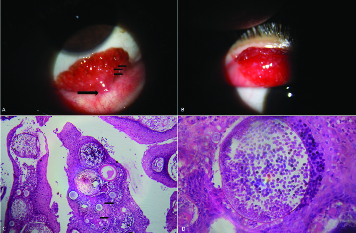
5)Clinical Features

The most common clinical presentation of conjunctival rhinosporidiosis includes:
- Red, fleshy, polypoidal mass on the conjunctiva
- Often appears pedunculated or sessile
- May be bleeding or covered with white dots (representing sporangia)
- Unilateral involvement is more common
- No pain, but patients may report:
◦ Foreign body sensation
◦ Mild irritation
◦ Epiphora (watering of eyes)
◦ Visual obstruction (if lesion is large)
Sites of involvement:
- Palpebral conjunctiva (especially tarsal)
- Bulbar conjunctiva
- Fornices
- Caruncle
- Lacrimal sac (less commonly)
6)Diagnosis
1. Clinical Examination
An ophthalmologist can suspect the diagnosis based on the appearance of a red, vascular, pedunculated mass with white dots.
2. Histopathology (Gold standard)
- Hematoxylin and eosin (H&E) staining reveals:
◦ Mature and immature sporangia
◦ Endospores within the sporangia
◦ Chronic inflammatory infiltrate
3. Special Stains
- Periodic acid-Schiff (PAS) and Gomori methenamine silver (GMS) stains highlight the wall of the sporangia.
4. Molecular Techniques
Rarely used in routine practice but helpful in research settings — PCR and rDNA sequencing have confirmed classification under Mesomycetozoea.
Differential Diagnosis
- Pyogenic granuloma
- Conjunctival papilloma
- Hemangioma
- Squamous cell carcinoma (especially in older patients)
- Kaposi sarcoma (in HIV patients)
- Ocular cysticercosis
Histopathology helps to distinguish rhinosporidiosis from these entities.
DISCLAIMER-Some patients go to net and directly take treatment from there which can lead to catastrophic consequences-Then- Many people ask then why to read all this text -the reason is that it helps you to understand the pathology better ,you can cooperate with treatment better ,your treating physician is already busy with his patients and he does not have sufficient time to explain you all the things right from ABCD ,so it is always better to have some knowledge of the disease /disorder you are suffering from.
7)Management
1. Surgical Excision
- Complete excision of the lesion is the treatment of choice.
- Base of the lesion should be cauterized or treated with electrocoagulation to reduce recurrence.
- Great care must be taken to avoid rupture of sporangia during surgery to prevent autoinoculation or recurrence.

2. Medical Therapy
- Rhinosporidium seeberi is resistant to most antifungal and antibacterial drugs.
- Dapsone (100 mg/day for several months) has been used as an adjunct to:
◦ Arrest maturation of sporangia
◦ Promote fibrosis
◦ Reduce recurrence
However, its efficacy remains controversial and not universally accepted.
8)Complications
- Recurrence of the lesion (due to incomplete excision or autoinoculation)
- Scarring or fibrosis of conjunctiva
- Cosmetic disfigurement
- Rarely, spread to:
◦ Lacrimal sac (causing dacryocystitis)
◦ Orbit (causing proptosis)
◦ Nasal cavity (if conjunctival lesion extends)
9)Prevention
- Avoid bathing in contaminated or stagnant water, especially in endemic regions
- Maintain good ocular hygiene
- Do not rub eyes with unclean hands or cloths
- Community education in endemic regions about safe water practices
10)Prognosis
- With complete surgical excision, prognosis is generally good.
- Recurrence rate can vary between 10-25%, depending on surgical technique.
- Ocular rhinosporidiosis is non-fatal and does not usually impair vision unless complications arise.
11)Conclusion
Conjunctival rhinosporidiosis, though rare, should be considered in the differential diagnosis of conjunctival masses in patients from endemic areas or with a history of bathing in stagnant water. Accurate clinical recognition and complete surgical excision are the keys to effective treatment. Continued public health awareness and research are necessary to better understand and manage this unusual but significant ocular infection.
FOR FURTHER INFORMATION IN GREAT DETAIL ON Rhinosporidiosis PL CLICK ON THE LINK GIVEN BELOW-It is always better to view links from laptop/desktop rather than mobile phone as they may not be seen from mobile phone. ,in case of technical difficulties you need to copy paste this link in google search. In case if you are viewing this blog from mobile phone you need to click on the three dots on the right upper corner of your mobile screen and ENABLE DESKTOP VERSION.
FOR INFORMATION IN GREAT DETAIL ON Rhinosporidiosis Pathology Outline PL CLICK ON THE LINK GIVEN BELOW-It is always better to view links from laptop/desktop rather than mobile phone as they may not be seen from mobile phone. ,in case of technical difficulties you need to copy paste this link in google search. In case if you are viewing this blog from mobile phone you need to click on the three dots on the right upper corner of your mobile screen and ENABLE DESKTOP VERSION.
FOR INFORMATION IN GREAT DETAIL ON Rhinosporidiosis Treatment PL CLICK ON THE LINK GIVEN BELOW-It is always better to view links from laptop/desktop rather than mobile phone as they may not be seen from mobile phone. ,in case of technical difficulties you need to copy paste this link in google search. In case if you are viewing this blog from mobile phone you need to click on the three dots on the right upper corner of your mobile screen and ENABLE DESKTOP VERSION.
FOR INFORMATION IN GREAT DETAIL ON Rhinosporidiosis Symptoms PL CLICK ON THE LINK GIVEN BELOW-It is always better to view links from laptop/desktop rather than mobile phone as they may not be seen from mobile phone. ,in case of technical difficulties you need to copy paste this link in google search. In case if you are viewing this blog from mobile phone you need to click on the three dots on the right upper corner of your mobile screen and ENABLE DESKTOP VERSION.
FOR FURTHER INFORMATION IN GREAT DETAIL ON Rhinosporidiosis Histology PL CLICK ON THE LINK GIVEN BELOW-It is always better to view links from laptop/desktop rather than mobile phone as they may not be seen from mobile phone. ,in case of technical difficulties you need to copy paste this link in google search. In case if you are viewing this blog from mobile phone you need to click on the three dots on the right upper corner of your mobile screen and ENABLE DESKTOP VERSION.
FOR FURTHER INFORMATION IN GREAT DETAIL ON Rhinosporidiosis Life Cycle PL CLICK ON THE LINK GIVEN BELOW-It is always better to view links from laptop/desktop rather than mobile phone as they may not be seen from mobile phone. ,in case of technical difficulties you need to copy paste this link in google search. In case if you are viewing this blog from mobile phone you need to click on the three dots on the right upper corner of your mobile screen and ENABLE DESKTOP VERSION.
FOR FURTHER INFORMATION IN GREAT DETAIL ON Rhinoscleroma vs Rhinosporidiosis PL CLICK ON THE LINK GIVEN BELOW-It is always better to view links from laptop/desktop rather than mobile phone as they may not be seen from mobile phone. ,in case of technical difficulties you need to copy paste this link in google search. In case if you are viewing this blog from mobile phone you need to click on the three dots on the right upper corner of your mobile screen and ENABLE DESKTOP VERSION.
FOR FURTHER INFORMATION IN GREAT DETAIL ON Ocular Rhinosporidiosis PL CLICK ON THE LINK GIVEN BELOW-It is always better to view links from laptop/desktop rather than mobile phone as they may not be seen from mobile phone. ,in case of technical difficulties you need to copy paste this link in google search. In case if you are viewing this blog from mobile phone you need to click on the three dots on the right upper corner of your mobile screen and ENABLE DESKTOP VERSION.
If any patient has any ENT -Ear nose throat problems and requires any , consultation ,online consultation ,or surgery in clinic of ENT specialist Doctor Dr Sagar Rajkuwar ,he may TAKE APPOINTMENT BY CLICKING ON THE LINK GIVEN BELOW-
Clinic address of ENT SPECIALIST doctor Dr Sagar Rajkuwar-
Prabha ENT clinic, plot no 345,Saigram colony, opposite Indoline furniture Ambad link road ,Ambad ,1 km from Pathardi phata Nashik ,422010 ,Maharashtra, India-Dr Sagar Rajkuwar (MS-ENT), Cel no- 7387590194 , 9892596635


