Rhinosporidiosis Histology-various aspects-
Rhinosporidiosis Histology: A Comprehensive Overview
Introduction
Rhinosporidiosis is a chronic granulomatous disease caused by the pathogen Rhinosporidium seeberi, which primarily affects the mucous membranes of the nasal cavity, conjunctiva, and other sites. Although traditionally considered a fungus, molecular studies have shown that Rhinosporidium seeberi belongs to a novel group of aquatic protists in the class Mesomycetozoea, closely related to fish pathogens.


Histological examination remains the gold standard for diagnosis, as the organism cannot be cultured in vitro. The disease is characterized histologically by the presence of large, thick-walled sporangia containing endospores in a granulomatous inflammatory background.
If Any Patient of ENT Requires Any Surgery, Opd Consultation Or Online Consultation In Clinic of ENT Specialist Doctor Dr. Sagar Rajkuwar ,He May Contact Him At The Following Address-
Prabha ENT Clinic, Plot no 345,Saigram Colony, Opposite Indoline Furniture Ambad Link Road ,Ambad ,1 km From Pathardi Phata Nashik ,422010 ,Maharashtra, India-Dr. Sagar Rajkuwar (MS-ENT), Cell No- 7387590194, 9892596635
Etiology and Pathogenesis
Rhinosporidium seeberi is an aquatic protist. It is hypothesized to enter the body via traumatized epithelium, most commonly in individuals exposed to stagnant water (ponds, lakes, etc.). The nasal mucosa is the most frequent site of entry and colonization, although other mucosal sites such as the conjunctiva, larynx, trachea, skin, and even genitalia can be involved.
Once inside the host tissue, the organism undergoes a complex developmental cycle, forming large, mature sporangia. The sporangia eventually rupture, releasing endospores that can initiate further infection in adjacent tissue.
Gross Pathology
The lesions of rhinosporidiosis typically present as polypoidal, friable, and vascular masses, often with a characteristic strawberry-like appearance due to the presence of visible white dots (sporangia) on the surface. They bleed easily on manipulation or trauma.
Microscopic (Histological) Features
Histologically, rhinosporidiosis has several pathognomonic features, which can be identified using Hematoxylin and Eosin (H&E) staining and special stains such as Periodic acid–Schiff (PAS) or Gomori methenamine silver (GMS).
1. Sporangia
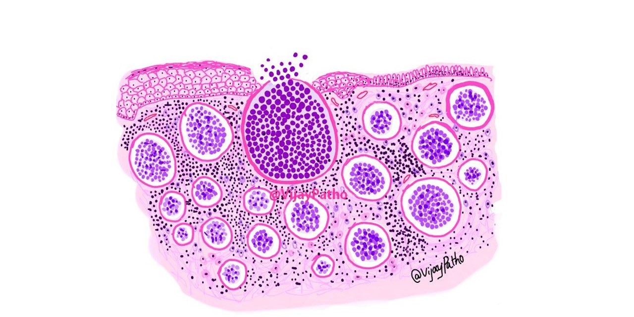
- The hallmark of rhinosporidiosis is the presence of large, thick-walled sporangia embedded in the subepithelial tissue.
- These sporangia can range in size from 10 microns to over 350 microns in diameter.
- Each sporangium has a triple-layered chitinous wall, which stains positively with PAS and GMS.
- The interior of the mature sporangia contains hundreds of endospores, which are spherical to oval, measure around 5–10 microns, and are usually basophilic.
2. Stages of Sporangial Maturation
Histologically, sporangia can be seen in different stages of development:
- Young trophic stages: Small basophilic bodies without a defined wall, embedded in the host tissue.
- Immature sporangia: Intermediate size with partial wall formation and some internal division.
- Mature sporangia: Large with well-formed triple-layered walls and densely packed with endospores.
- Ruptured sporangia: These may release endospores into the surrounding tissue, inciting an inflammatory response.
For Update On Further Important Health Related Topics And Frequently Asked Questions On Health Topics By General Population Please Click On The Link Given Below To Join Our WhatsApp Group –
https://chat.whatsapp.com/Lv3NbcguOBS5ow6X9DpMMA
3. Inflammatory Response
- The surrounding tissue shows a mixed inflammatory infiltrate consisting of:
◦ Lymphocytes
◦ Plasma cells
◦ Neutrophils
◦ Occasional eosinophils
◦ Foreign body-type multinucleated giant cells surrounding ruptured sporangia - Granulomatous inflammation may be seen, especially in chronic lesions.
- Fibrosis may develop around older lesions.


4. Epithelial Changes
- The overlying epithelium may show hyperplasia, ulceration, or even pseudoepitheliomatous hyperplasia.
- Surface ulceration often occurs due to the mechanical stress or rupture of superficial sporangia.
Differential Diagnosis (Histologically)
Histologically, rhinosporidiosis can be confused with several conditions. However, the size, morphology, and staining characteristics of the sporangia help differentiate it from other entities.
DISCLAIMER-Some patients go to net and directly take treatment from there which can lead to catastrophic consequences-Then- Many people ask then why to read all this text -the reason is that it helps you to understand the pathology better ,you can cooperate with treatment better ,your treating physician is already busy with his patients and he does not have sufficient time to explain you all the things right from ABCD ,so it is always better to have some knowledge of the disease /disorder you are suffering from.
- Coccidioidomycosis:
◦ Caused by Coccidioides immitis, which also forms spherules with endospores.
◦ Spherules are smaller (20–80 microns) and have thinner walls.
◦ Requires correlation with clinical and geographic history. - Myospherulosis:
◦ Caused by lipid-rich materials; shows pseudocystic spaces with spherule-like structures.
◦ No true sporangia or endospores; often a reaction to petroleum-based substances. - Cryptococcosis:
◦ Caused by Cryptococcus neoformans, smaller yeast with narrow-based budding and mucicarmine-positive capsule. - Paracoccidioidomycosis:
◦ Shows “pilot wheel” morphology with multiple buds; not seen in rhinosporidiosis.
Special Stains and Diagnostic Tools
- PAS: Stains the sporangial wall and endospores magenta; highlights the presence of polysaccharides.
- GMS: Stains the sporangial wall black; useful in differentiating from other fungi.
- Mucicarmine: Typically negative in rhinosporidiosis (unlike cryptococcosis).
- Immunohistochemistry: Rarely used; no specific markers are widely available.
Molecular Studies
Modern molecular techniques have placed Rhinosporidium seeberi within the Mesomycetozoea class of aquatic protists. However, molecular diagnostics are not routinely available due to the lack of widespread PCR-based kits or specific probes.
Clinical Correlation
Histological findings must always be interpreted in conjunction with clinical features. Most patients present with:
- Nasal obstruction
- Epistaxis
- Polyps in nasal or ocular mucosa
The diagnosis is often confirmed after biopsy or excision of the lesion and histopathological examination.
Treatment and Prognosis
There is no effective medical therapy due to the inability to culture the organism and test susceptibility. Surgical excision with wide margins and electrocauterization of the base is the treatment of choice. Recurrence is common due to:
- Incomplete excision
- Release of endospores during surgery
Conclusion
Histology is critical in diagnosing rhinosporidiosis, especially given the organism’s inability to be cultured in vitro. The characteristic appearance of large sporangia with endospores in an inflammatory stroma is diagnostic. Awareness of the histopathological features helps distinguish rhinosporidiosis from other granulomatous infections and fungal diseases.
Continued research into molecular diagnostics and epidemiology will further refine our understanding and management of this unique infectious disease.
FOR FURTHER INFORMATION IN GREAT DETAIL ON Rhinosporidiosis PL CLICK ON THE LINK GIVEN BELOW-It is always better to view links from laptop/desktop rather than mobile phone as they may not be seen from mobile phone. ,in case of technical difficulties you need to copy paste this link in google search. In case if you are viewing this blog from mobile phone you need to click on the three dots on the right upper corner of your mobile screen and ENABLE DESKTOP VERSION.
FOR INFORMATION IN GREAT DETAIL ON Rhinosporidiosis Pathology Outline PL CLICK ON THE LINK GIVEN BELOW-It is always better to view links from laptop/desktop rather than mobile phone as they may not be seen from mobile phone. ,in case of technical difficulties you need to copy paste this link in google search. In case if you are viewing this blog from mobile phone you need to click on the three dots on the right upper corner of your mobile screen and ENABLE DESKTOP VERSION.
FOR INFORMATION IN GREAT DETAIL ON Rhinosporidiosis Treatment PL CLICK ON THE LINK GIVEN BELOW-It is always better to view links from laptop/desktop rather than mobile phone as they may not be seen from mobile phone. ,in case of technical difficulties you need to copy paste this link in google search. In case if you are viewing this blog from mobile phone you need to click on the three dots on the right upper corner of your mobile screen and ENABLE DESKTOP VERSION.
FOR INFORMATION IN GREAT DETAIL ON Rhinosporidiosis Symptoms PL CLICK ON THE LINK GIVEN BELOW-It is always better to view links from laptop/desktop rather than mobile phone as they may not be seen from mobile phone. ,in case of technical difficulties you need to copy paste this link in google search. In case if you are viewing this blog from mobile phone you need to click on the three dots on the right upper corner of your mobile screen and ENABLE DESKTOP VERSION.
If any patient has any ENT -Ear nose throat problems and requires any , consultation ,online consultation ,or surgery in clinic of ENT specialist Doctor Dr Sagar Rajkuwar ,he may TAKE APPOINTMENT BY CLICKING ON THE LINK GIVEN BELOW-
Clinic address of ENT SPECIALIST doctor Dr Sagar Rajkuwar-
Prabha ENT clinic, plot no 345,Saigram colony, opposite Indoline furniture Ambad link road ,Ambad ,1 km from Pathardi phata Nashik ,422010 ,Maharashtra, India-Dr Sagar Rajkuwar (MS-ENT), Cel no- 7387590194 , 9892596635


