Sinus lift surgery -various aspects-
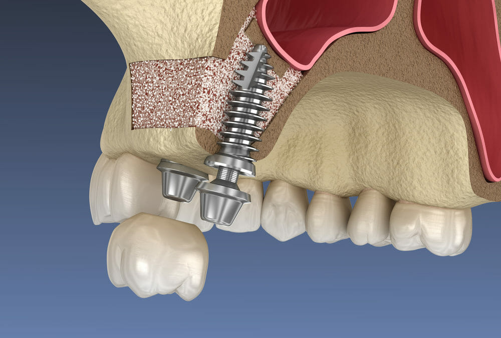
A sinus lift is a Surgical operation that enhances the quantity of bone in the upper jaw to support dental implants. It is also referred to as sinus augmentation.


Why it is conducted
When there is inadequate bone in the upper jaw to support dental implants
When the sinuses are positioned too closely to the jaw
How it is conducted
The sinus membrane is elevated
A bone graft is inserted between the jaw and the sinus membrane
The bone graft integrates with the jaw
Dental implants are inserted into the jaw
Risks and complications: Swelling, Hematoma, Perforation of the Schneiderian membrane, and Chronic rhinosinusitis.
After surgery
You may encounter some discomfort, swelling, or light bleeding from your nose or mouth
Avoid blowing your nose or sneezing often
Your dentist may provide you with saline spray to keep your nose hydrated
Cost
The expense varies based on the complexity of the procedure and the quantity of bone graft material required.
The crucial factor in ensuring the success of an implant lies in both the quantity and quality of the bone at the placement site. Historically, inserting dental implants in the upper posterior jaw has been challenging due to limited bone volume and density, along with the nearby sinus being in close proximity.
If Any Patient of ENT Requires Any Surgery, Opd Consultation Or Online Consultation In Clinic of ENT Specialist Doctor Dr. Sagar Rajkuwar ,He May Contact Him At The Following Address-
Prabha ENT Clinic, Plot no 345,Saigram Colony, Opposite Indoline Furniture Ambad Link Road ,Ambad ,1 km From Pathardi Phata Nashik ,422010 ,Maharashtra, India-Dr. Sagar Rajkuwar (MS-ENT), Cell No- 7387590194, 9892596635
Why Sinus lift surgery is required ?
In cases where bone loss has occurred in that specific area, possibly due to factors like periodontal disease or teeth loss, there might not be sufficient bone available for implant placement.
Sinus lift surgery, also referred to as sinus augmentation, is a procedure that can address this issue by elevating the sinus floor and creating bone for the insertion of dental implants. Various methods are available to elevate the sinus and promote the regeneration of new bone tissue.
Proceedure of sinus lift surgery –
In a typical procedure, a small cut is performed in the gum to reveal the underlying bone. Next, a small circle is carefully cut into the bone. With a gentle lift, this bony piece is placed into the sinus cavity, resembling a trap door, and then filled with bone graft material below. Your periodontist will be able to discuss with you the various bone graft materials available, which are designed to restore lost bone and tissue.
Recovery and Risks Recovery and Risks of sinus lift surgery –
Recovery typically takes several months, as the bone graft integrates with the existing bone.
Risks are generally low but can include infection, sinus problems, or graft rejection, though these complications are rare.
Sinus lift surgery is a crucial step for many people who need dental implants in the upper jaw and can dramatically improve the chances of successful implant placement .
Anatomy of the maxillary sinus in relation to sinus lift surgery –


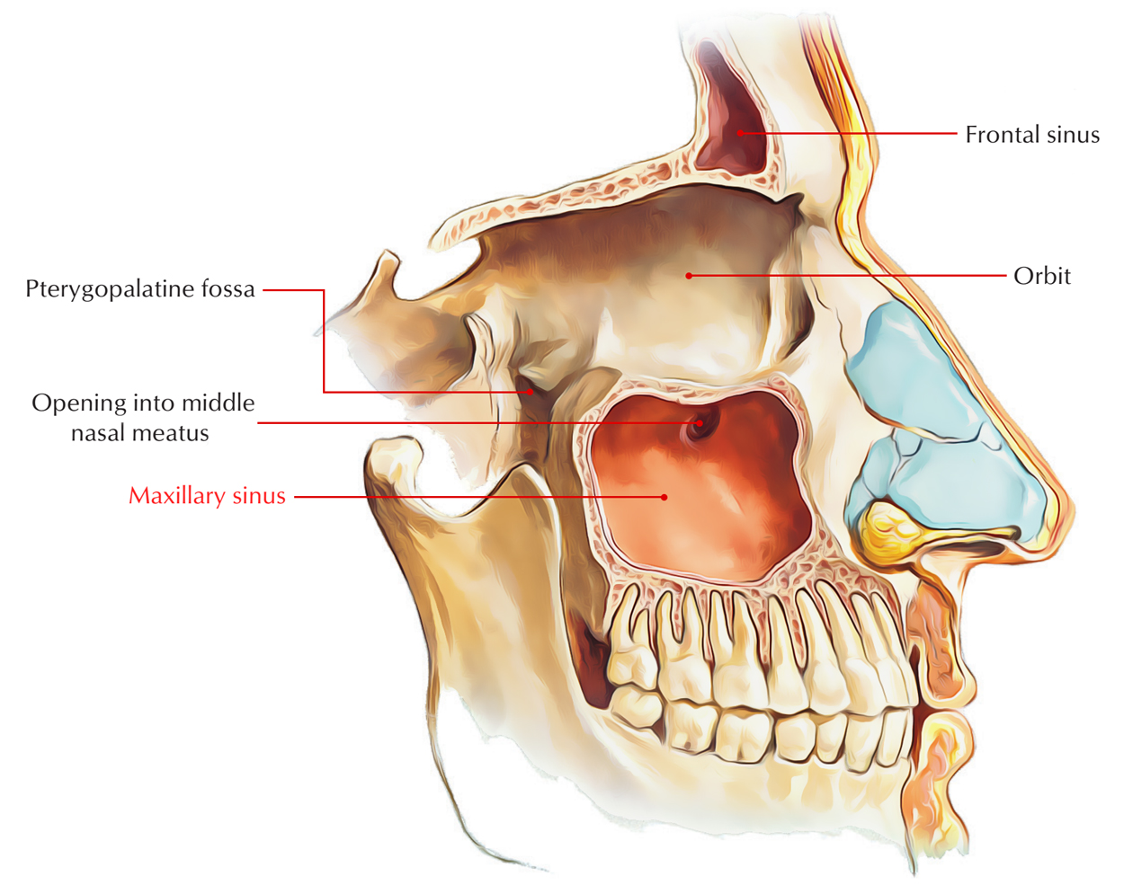
The maxillary sinus is an air-filled space that occupies the maxilla on both sides1 and is encircled by the nasal cavity in a medial direction, the maxillary tuberosity in a lateral direction, the orbit above, and the alveolar bone below.
Anatomy and Physiology of the Maxillary Sinus
The maxillary sinus, which is also referred to as the antrum of Highmore, is nearly absent in newborns. The maxillary sinus gradually fills with air over time, resulting in an increase in sinus volume as one ages. This pneumatization process continues for the duration of life, leading to the continually enlarging sinus cavity. The bone that is resorbed due to this cavity widening is the maxillary alveolar bone, which provides support to the teeth. The maxillary sinus is recognized as the largest of the paranasal sinuses. Its exact function is not clearly understood; however, it is believed to lighten the skull, humidify the air taken in, and assist in voice resonance. 3
The sinus
is lined with a pseudostratified ciliated columnar/cuboidal epithelium, referred to as the schneiderian membrane. This delicate membrane generates mucus through goblet cells and includes a basement membrane with sporadic osteoblasts. The ciliated membrane serves to transport mucus and debris to the ostium semilunaris, facilitating its exit from the sinus cavity. The ostium is located above the depth of the sinus floor, thus necessitating that the ciliated cells propel the mucus in a cephalad direction. The ostium is positioned within the semilunar hiatus of the middle meatus of the nasal cavity and is a narrow opening that can be easily blocked by mucosal swelling, consequently hindering proper drainage of the maxillary sinus.
Blood supply to the maxillary sinus is plentiful and comes from the following sources-FOR FURTHER INFORMATION IN GREAT DETAIL PL CLICK ON THE LINK GIVEN BELOW-It is always better to view links from laptop/desktop rather than mobile phone as they may not be seen from mobile phone. ,in case of technical difficulties you need to copy paste this link in google search. In case if you are viewing this blog from mobile phone you need to click on the three dots on the right upper corner of your mobile screen and ENABLE DESKTOP VERSION .
How can a Sinus lift procedure assist?
Numerous individuals who have lost their back teeth in the upper jaw lack sufficient bone for implant placement. Sinus lift procedures provide the opportunity to generate adequate bone beneath the sinus to accommodate dental implants in the back part of the upper jaw. The bone is introduced between your jaw and the sinuses. The duration from this procedure to dental restoration can range from six to twelve months but may extend longer.
What varieties of sinus lift procedures are accessible?


There are two primary categories of procedures based on the quantity of existing bone at the intended implant site:
1a) External approach without existing bone
Grafting material will be applied from the cheek side of your sinus to elevate the membrane. The dental implant is typically not placed until the bone has completely healed, necessitating an additional surgical procedure for this.
1b) External approach with some existing bone
Grafting material will be applied from the cheek side of your sinus to elevate the membrane. Your dental implant can be positioned simultaneously.
2) Internal approach with some existing bone
Your sinus will be elevated by gently tapping through the implant preparation site in your mouth. Grafting material may be inserted through the implant preparation site, allowing your dental implant to be placed concurrently.
What grafting materials are utilized in sinus lift procedures?
Bone grafting materials sourced from your body, other human sources, animals, and synthetic origins are available. The graft material forms a framework for your own bone to develop into. This process could take four to twelve months.
The most frequently used material in sinus lifts is referred to as Bio-Oss. This artificial sterilized bone is derived from cattle and is processed for safety in human applications. Moreover, a membrane patch might be positioned over the bone graft for its protection.
The most commonly utilized membrane for sinus lift is Bio-Gide, which is absorbable. This artificial membrane is derived from porcine (pig) sources. Your surgeon will clarify which products they plan to use for your sinus lift.
If you have any reservations regarding the use of animal-derived products, please bring them up with your surgeon.
What are the potential risks associated with sinus lift procedures?
In typical healing, you should anticipate:
pain to be managed with basic pain relievers such as ibuprofen and paracetamol
swelling to peak two days post-procedure
bruising around the cheek expanding over the lower jaw
an improvement in your condition after stitch removal in ten days
a return to normalcy after two to three weeks.
Is a sinus lift a serious surgery?
How painful is a sinus lift?
Prabha ENT Clinic, Plot no 345,Saigram Colony, Opposite Indoline Furniture Ambad Link Road ,Ambad ,1 km From Pathardi Phata Nashik ,422010 ,Maharashtra, India-Dr. Sagar Rajkuwar (MS-ENT), Cell No- 7387590194, 9892596635
Issued in public interest by –
www.entspecialistinnashik.com


