Rhinosporidiosis Symptoms-various aspects-
Rhinosporidiosis Symptoms – A Comprehensive Overview
Introduction
Rhinosporidiosis is a chronic granulomatous infection caused by the aquatic protist Rhinosporidium seeberi, affecting mucous membranes. The disease is characterized by the formation of highly vascular, polypoidal, friable lesions which bleed easily and have a tendency to recur.

The infection can affect various parts of the body, and its symptoms vary depending on the site of involvement. Although the nose and nasopharynx are the most commonly affected areas, other anatomical regions such as the ocular structures, skin, genitalia, and in rare cases, visceral organs can also be involved.
This comprehensive guide will cover the range of symptoms associated with rhinosporidiosis, providing detailed descriptions based on the location of the lesion, its appearance, progression, complications, and differentiating features from similar conditions.
If Any Patient of ENT Requires Any Surgery, Opd Consultation Or Online Consultation In Clinic of ENT Specialist Doctor Dr. Sagar Rajkuwar ,He May Contact Him At The Following Address-
Prabha ENT Clinic, Plot no 345,Saigram Colony, Opposite Indoline Furniture Ambad Link Road ,Ambad ,1 km From Pathardi Phata Nashik ,422010 ,Maharashtra, India-Dr. Sagar Rajkuwar (MS-ENT), Cell No- 7387590194, 9892596635
General Characteristics of Rhinosporidiosis Lesions
Before delving into site-specific symptoms, understanding the general properties of rhinosporidial lesions helps appreciate their clinical manifestations:
- Polypoid or nodular growths
- Highly vascularized and friable (bleed easily)
- Sessile or pedunculated
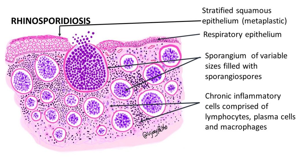
- Surface studded with white dots – these represent sporangia containing endospores
- Painless, but can lead to discomfort due to mass effect
- Slow-growing but persistent
- Often recurrent after removal
1. Nasal and Nasopharyngeal Rhinosporidiosis
This is the most common form, accounting for over 70% of cases. Patients typically present with:
Key Symptoms:
-
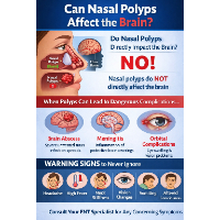
Nasal obstruction:
◦ Unilateral or bilateral, depending on lesion size
◦ Progressive blockage over weeks or months -
Nasal discharge:
◦ Mucoid, mucopurulent, or blood-tinged
◦ Often chronic and associated with crusting
-
Frequent epistaxis (nosebleeds):
◦ Occurs due to the high vascularity of the lesion
◦ Can be spontaneous or triggered by touch
-
Visible nasal mass:
◦ Red, fleshy, polyp-like growth seen on anterior rhinoscopy
◦ Often confused with ordinary nasal polyps, but rhinosporidial polyps are more vascular
-
Sneezing and nasal irritation:
◦ Secondary to mucosal inflammation
-
Hyposmia or anosmia:
◦ Loss of smell in cases of extensive nasal blockage
-
Foreign body sensation:
◦ Due to lesion size and location, especially when located in posterior nasal cavity
-
Change in voice:
◦ Nasal speech if the mass obstructs airflow
-
Mouth breathing:
◦ Common in children with nasal obstruction
For Update On Further Important Health Related Topics And Frequently Asked Questions On Health Topics By General Population Please Click On The Link Given Below To Join Our WhatsApp Group –
https://chat.whatsapp.com/Lv3NbcguOBS5ow6X9DpMMA
Advanced Cases May Show:
- Extension into the oropharynx or nasopharynx, causing difficulty in breathing or swallowing
- Facial deformity or external swelling over the nasal bridge in long-standing, untreated cases
2. Ocular Rhinosporidiosis
Also known as oculo-rhinosporidiosis, this is the second most common presentation, often affecting the conjunctiva or lacrimal sac.

Symptoms by Ocular Subsite:
a. Conjunctiva:
- Red, lobulated mass on the bulbar or palpebral conjunctiva
- Epiphora (excessive tearing)
- Recurrent conjunctivitis-like symptoms
- Irritation or foreign body sensation
- Bleeding from eye on rubbing
- Visual disturbances, especially if mass encroaches on cornea
b. Lacrimal sac:
- Swelling near the medial canthus
- Mucoid or bloody discharge from punctum
- May mimic chronic dacryocystitis
Differentiating Features:
- Presence of granular surface with sporangial dots
- Recurrent growths despite excision
3. Cutaneous Rhinosporidiosis
Cutaneous involvement may be primary (through inoculation) or secondary (via autoinoculation or dissemination). It presents with a wide range of symptoms depending on depth and duration.
Cutaneous Symptoms:
-
Nodular or verrucous growths:
◦ Red to violaceous nodules
◦ May resemble viral warts or squamous papillomas
-
Ulcerative lesions:
◦ Especially if the lesion gets traumatized or secondarily infected
-
Plaque-like formations:
◦ Resemble chronic granulomatous diseases like lupus vulgaris
-
Pedunculated masses:
◦ Mimic pyogenic granulomas
- Localized swelling with draining sinuses
- Painless or mildly painful
- Recurrent lesions after excision
Cutaneous lesions often occur near previously infected mucosal areas or at trauma-prone sites such as the face, chest, back, or limbs.
DISCLAIMER-Some patients go to net and directly take treatment from there which can lead to catastrophic consequences-Then- Many people ask then why to read all this text -the reason is that it helps you to understand the pathology better ,you can cooperate with treatment better ,your treating physician is already busy with his patients and he does not have sufficient time to explain you all the things right from ABCD ,so it is always better to have some knowledge of the disease /disorder you are suffering from.
4. Genitourinary Rhinosporidiosis
Although rare, involvement of genital and perineal regions is documented.
In Males:
- Lesions on the penis, scrotum, or urethral meatus
- Dysuria, hematuria, or urinary obstruction if the urethra is involved
- May cause pain during urination or intercourse
In Females:
- Involvement of the vagina, cervix, or vulva
- May mimic genital warts or carcinomas
5. Disseminated Rhinosporidiosis
This is extremely rare and mostly affects immunocompromised individuals or those with long-standing untreated disease.
Symptoms Based on Site:
Lungs:
- Chronic cough
- Hemoptysis (coughing up blood)
- Chest discomfort
Bones:
- Swelling and tenderness over bones
- Radiological evidence of osteolytic lesions
- Pathological fractures
Viscera (liver, spleen, etc.):
- Organomegaly
- Abdominal pain or mass
- Constitutional symptoms like fever and weight loss
Disseminated disease may involve multiple organs simultaneously and requires high clinical suspicion.
Complications Due to Symptoms
If not properly treated, rhinosporidiosis can lead to several complications:
- Secondary bacterial infections in ulcerated lesions
- Airway obstruction from large nasopharyngeal growths
- Vision impairment in ocular cases
- Cosmetic disfigurement of face or external genitalia
- Psychological distress due to recurrence or chronicity
Natural Progression and Recurrence
Rhinosporidial lesions often progress slowly over months or years. In the early stages, the symptoms may be mild, but they worsen over time as the lesion grows in size. Surgical excision is the mainstay of treatment, but recurrence is common due to:
- Incomplete removal
- Intraoperative rupture and spillage of endospores
- Lack of cauterization of the lesion base
Symptoms often recur at the same site or, less commonly, appear at a new site due to autoinoculation.
Symptom-Based Clues to Diagnosis
Some characteristic signs and symptoms that suggest rhinosporidiosis over other conditions:
- Red, bleeding nasal or conjunctival mass with white dots
- Recurrent lesions after multiple surgical removals
- Polyp-like masses in people from endemic areas with history of bathing in ponds or rivers
- Nasal obstruction with epistaxis in absence of other nasal disease
- Lacrimal sac swelling with mucoid discharge unresponsive to antibiotics
Conclusion
The symptoms of rhinosporidiosis vary significantly depending on the affected organ, stage of disease, and extent of spread. From nasal obstruction and epistaxis to conjunctival masses, cutaneous nodules, and systemic involvement, the spectrum of presentation is wide. Recognizing these symptoms early, particularly in endemic areas, is critical for accurate diagnosis and timely treatment.
Since the disease can recur frequently, a multidisciplinary approach combining ENT specialists, dermatologists, ophthalmologists, and surgeons is often needed to manage symptoms effectively and minimize morbidity.
FOR FURTHER INFORMATION IN GREAT DETAIL ON Rhinosporidiosis PL CLICK ON THE LINK GIVEN BELOW-It is always better to view links from laptop/desktop rather than mobile phone as they may not be seen from mobile phone. ,in case of technical difficulties you need to copy paste this link in google search. In case if you are viewing this blog from mobile phone you need to click on the three dots on the right upper corner of your mobile screen and ENABLE DESKTOP VERSION.
FOR INFORMATION IN GREAT DETAIL ON Rhinosporidiosis Pathology Outline PL CLICK ON THE LINK GIVEN BELOW-It is always better to view links from laptop/desktop rather than mobile phone as they may not be seen from mobile phone. ,in case of technical difficulties you need to copy paste this link in google search. In case if you are viewing this blog from mobile phone you need to click on the three dots on the right upper corner of your mobile screen and ENABLE DESKTOP VERSION.
FOR INFORMATION IN GREAT DETAIL ON Rhinosporidiosis Treatment PL CLICK ON THE LINK GIVEN BELOW-It is always better to view links from laptop/desktop rather than mobile phone as they may not be seen from mobile phone. ,in case of technical difficulties you need to copy paste this link in google search. In case if you are viewing this blog from mobile phone you need to click on the three dots on the right upper corner of your mobile screen and ENABLE DESKTOP VERSION.
If any patient has any ENT -Ear nose throat problems and requires any , consultation ,online consultation ,or surgery in clinic of ENT specialist Doctor Dr Sagar Rajkuwar ,he may TAKE APPOINTMENT BY CLICKING ON THE LINK GIVEN BELOW-
Clinic address of ENT SPECIALIST doctor Dr Sagar Rajkuwar-
Prabha ENT clinic, plot no 345,Saigram colony, opposite Indoline furniture Ambad link road ,Ambad ,1 km from Pathardi phata Nashik ,422010 ,Maharashtra, India-Dr Sagar Rajkuwar (MS-ENT), Cel no- 7387590194 , 9892596635