Rhinosporidiosis Pathology Outline -various aspects-
I. Introduction
Rhinosporidiosis is a chronic granulomatous disease caused by the eukaryotic pathogen Rhinosporidium seeberi. Though previously thought to be a fungus, it is now classified under Mesomycetozoea, a group of aquatic protists. The hallmark of rhinosporidiosis is polypoidal mucosal lesions, predominantly affecting the nasal mucosa, with characteristic histopathological features including large sporangia containing numerous endospores.

It is a non-contagious disease that primarily affects humans and animals exposed to stagnant water and is endemic in India, Sri Lanka, and parts of Africa and South America.
II. Gross Pathology
A. Site of Lesion
- Nasal cavity: Most common site (approximately 70–80% of cases)
- Other sites:
◦ Nasopharynx
◦ Conjunctiva and lacrimal sac (ocular involvement)
◦ Skin (cutaneous)
◦ Genitourinary tract (penis, urethra)
◦ Rarely, systemic sites (bone, lungs, brain)
If Any Patient of ENT Requires Any Surgery, Opd Consultation Or Online Consultation In Clinic of ENT Specialist Doctor Dr. Sagar Rajkuwar ,He May Contact Him At The Following Address-
Prabha ENT Clinic, Plot no 345,Saigram Colony, Opposite Indoline Furniture Ambad Link Road ,Ambad ,1 km From Pathardi Phata Nashik ,422010 ,Maharashtra, India-Dr. Sagar Rajkuwar (MS-ENT), Cell No- 7387590194, 9892596635
B. Gross Morphology
- Polypoidal, fleshy mass attached to the mucosa by a pedicle or broad base
- Color: Pink to reddish; surface may show tiny white dots representing mature sporangia
- Size: Varies from a few mm to several cm
- Consistency: Soft and friable
- Bleeds easily on touch or manipulation
- Surface may be ulcerated in recurrent or traumatized lesions
III. Microscopic Pathology (Histopathology)
Histopathology is the gold standard for diagnosis of rhinosporidiosis.
A. Epithelium
- Lined by hyperplastic stratified squamous or respiratory-type epithelium
- Often shows pseudoepitheliomatous hyperplasia (PEH)
- Foci of ulceration may be present
B. Subepithelial Tissue
- Dense granulomatous inflammation
- Prominent infiltrate with:
◦ Lymphocytes
◦ Plasma cells
◦ Histiocytes
◦ Multinucleated giant cells - Numerous sporangia of varying sizes are embedded within the stroma
C. Sporangia (Pathognomonic)
- Appear as thick-walled spherical to oval cysts, ranging from 50 to 400 microns
- Stages of development:
1. Immature sporangia: Smaller, with homogeneous eosinophilic contents
2. Mature sporangia: Larger, with visible internal endospores
3. Ruptured sporangia: Endospores released into surrounding tissue - Surrounded by a fibrous capsule and inflammatory cells
- Wall of sporangium: Refractile, eosinophilic, 5–10 µm thick
D. Endospores
- Small, round, basophilic bodies (5–10 µm)
- May be extracellular following rupture of the parent sporangium
- Some phagocytosed by giant cells and macrophages
E. Other Features
- Chronic inflammation with occasional microabscesses
- Fibrosis in long-standing lesions
- Secondary bacterial colonization may be present in ulcerated lesions
For Update On Further Important Health Related Topics And Frequently Asked Questions On Health Topics By General Population Please Click On The Link Given Below To Join Our WhatsApp Group –
https://chat.whatsapp.com/Lv3NbcguOBS5ow6X9DpMMA
IV. Stains and Special Techniques
A. Routine Stains
- Hematoxylin and Eosin (H&E):
◦ Sporangia seen as round to oval cystic structures with eosinophilic walls
◦ Endospores stain basophilic
◦ Surrounding granulomatous reaction is well visualized
![]()
B. Special Stains
- Periodic Acid-Schiff (PAS):
◦ Highlights the polysaccharide-rich wall of sporangia (magenta staining) - Gomori’s Methenamine Silver (GMS):
◦ Black staining of the sporangia and endospores - Mucicarmine: May also highlight the capsule
- Ziehl-Neelsen (ZN): Negative (helps rule out tuberculosis)
C. Immunohistochemistry and Molecular Techniques
- Not routinely used due to lack of standardized antibodies
- PCR for R. seeberi 18S rRNA gene has been developed in research settings
V. Cytology
A. Fine Needle Aspiration Cytology (FNAC)
- May reveal sporangia and endospores
- Background inflammatory infiltrate
- Helpful in diagnosis of cutaneous and subcutaneous lesions
B. Nasal Smear or Scrapings
- May demonstrate sporangia in superficial scrapings
- Less sensitive compared to biopsy
VI. Pathogenesis and Immune Response
A. Entry
- Through traumatized mucosa exposed to contaminated water
- Most likely via bathing or swimming in ponds/lakes
B. Sporangial Development
- The pathogen develops into mature sporangia within subepithelial tissue
- Each sporangium contains thousands of endospores
- Rupture leads to local spread and inflammation
DISCLAIMER-Some patients go to net and directly take treatment from there which can lead to catastrophic consequences-Then- Many people ask then why to read all this text -the reason is that it helps you to understand the pathology better ,you can cooperate with treatment better ,your treating physician is already busy with his patients and he does not have sufficient time to explain you all the things right from ABCD ,so it is always better to have some knowledge of the disease /disorder you are suffering from.
C. Host Response
- Mixed cellular immune response:
◦ Th1-type immunity associated with granulomatous response
◦ Delayed-type hypersensitivity may contribute to tissue damage - Failure to clear the organism results in chronic, recurrent lesions
VII. Differential Diagnosis (Histological)

- Fungal Infections
◦ Mucormycosis, aspergillosis, histoplasmosis
◦ Lack of characteristic large sporangia and endospores - Coccidioidomycosis
◦ May resemble rhinosporidiosis due to spherules with endospores
◦ Sporangia in coccidioidomycosis are smaller (30–60 µm) - Mycobacterial Infection (e.g., TB)
◦ Caseating granulomas and acid-fast bacilli (AFB-positive)
◦ No sporangia or endospores - Schistosomiasis
◦ May involve mucosa with granulomatous inflammation
◦ Presence of ova with terminal spine - Chronic Nasal Polyps or Inflammatory Polyps
◦ Lack the large sporangia or granulomatous component - Inverted Papilloma / Squamous Cell Carcinoma
◦ Pseudoepitheliomatous hyperplasia in rhinosporidiosis can mimic neoplasia
◦ Careful histological analysis is essential to avoid misdiagnosis
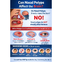
VIII. Complications (Pathological)
- Recurrent Lesions due to incomplete removal or residual spores
- Secondary bacterial infection in ulcerated tissues
- Fibrosis and scarring following repeated inflammation
- Obstruction (e.g., nasal airway, lacrimal duct, urethra)
- Extension into adjacent tissues: sinuses, pharynx, or skin
IX. Histopathological Classification (Proposed Staging)
Some researchers propose classification based on maturity of sporangia and tissue response:
- Early stage: Few immature sporangia, mild inflammation
- Mature stage: Large sporangia, dense mixed infiltrate
- Ruptured stage: Extracellular endospores, granulomas, fibrosis
X. Diagnostic Challenges and Pitfalls
- Misdiagnosed as fungal infection, neoplasia, or TB if pathognomonic features are missed
- PEH may mimic malignancy
- Unusual sites (e.g., skin, urethra) may delay diagnosis
- Need for adequate biopsy to visualize sporangia
FOR FURTHER INFORMATION IN GREAT DETAIL ON Rhinosporidiosis PL CLICK ON THE LINK GIVEN BELOW-It is always better to view links from laptop/desktop rather than mobile phone as they may not be seen from mobile phone. ,in case of technical difficulties you need to copy paste this link in google search. In case if you are viewing this blog from mobile phone you need to click on the three dots on the right upper corner of your mobile screen and ENABLE DESKTOP VERSION.
If any patient has any ENT -Ear nose throat problems and requires any , consultation ,online consultation ,or surgery in clinic of ENT specialist Doctor Dr Sagar Rajkuwar ,he may TAKE APPOINTMENT BY CLICKING ON THE LINK GIVEN BELOW-
Clinic address of ENT SPECIALIST doctor Dr Sagar Rajkuwar-
Prabha ENT clinic, plot no 345,Saigram colony, opposite Indoline furniture Ambad link road ,Ambad ,1 km from Pathardi phata Nashik ,422010 ,Maharashtra, India-Dr Sagar Rajkuwar (MS-ENT), Cel no- 7387590194 , 9892596635