Rhinosporidiosis Microscopy-various
Rhinosporidiosis Microscopy – A Detailed Overview
1. Introduction to Rhinosporidiosis
Rhinosporidiosis is a chronic granulomatous infection caused by the aquatic protist Rhinosporidium seeberi. It primarily affects the mucous membranes of the nasal cavity, nasopharynx, conjunctiva, and other parts of the upper respiratory tract. In rare cases, it can involve the skin, genitourinary tract, and other systemic organs.
This disease is endemic in parts of India (especially Tamil Nadu, Kerala, and Odisha), Sri Lanka, and parts of Africa and South America. Rhinosporidiosis is most common in individuals who frequently bathe in stagnant water bodies such as ponds or lakes, which are believed to harbor the infectious agent.
2. Causative Agent: Rhinosporidium seeberi
Rhinosporidium seeberi was originally classified as a fungus due to its spore-forming nature and staining characteristics. However, with advances in molecular biology and phylogenetic analysis, it is now classified as a protist, specifically under the class Mesomycetozoea, a group of aquatic protists that includes several other pathogens of fish and amphibians.
Despite extensive research, Rhinosporidium seeberi has never been successfully cultured in vitro, making its study primarily reliant on histopathology and molecular techniques.
If Any Patient of ENT Requires Any Surgery, Opd Consultation Or Online Consultation In Clinic of ENT Specialist Doctor Dr. Sagar Rajkuwar ,He May Contact Him At The Following Address-
Prabha ENT Clinic, Plot no 345,Saigram Colony, Opposite Indoline Furniture Ambad Link Road ,Ambad ,1 km From Pathardi Phata Nashik ,422010 ,Maharashtra, India-Dr. Sagar Rajkuwar (MS-ENT), Cell No- 7387590194, 9892596635
3. Pathogenesis
Infection occurs when the organism gains entry through traumatized epithelium, most commonly through the nasal mucosa. The exact mode of transmission is not fully understood, but contact with contaminated stagnant water is a well-established risk factor.
Once inside the host tissue, Rhinosporidium seeberi forms sporangia, which are large spherical structures that mature and eventually release endospores. These endospores can infect nearby tissue and continue the cycle.
4. Clinical Presentation
The classic clinical manifestation is a polypoidal, vascular, friable mass in the nasal cavity or nasopharynx. It may be mistaken clinically for nasal polyps or neoplasms. Other presentations include conjunctival growths (ocular rhinosporidiosis), cutaneous nodules, and rarely, disseminated disease in immunocompromised patients.
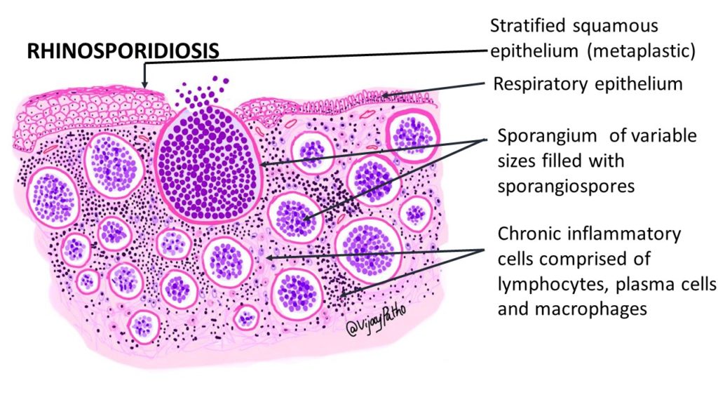
5. Microscopic Examination (Histopathology)
Microscopy remains the gold standard for diagnosis, given the organism’s inability to grow in culture. Examination of biopsied tissue provides a definitive diagnosis.
For Update On Further Important Health Related Topics And Frequently Asked Questions On Health Topics By General Population Please Click On The Link Given Below To Join Our WhatsApp Group –
https://chat.whatsapp.com/Lv3NbcguOBS5ow6X9DpMMA
Key Microscopic Features
1. Tissue Infiltration
- The tissue typically shows granulomatous inflammation with a mixture of chronic inflammatory cells, including lymphocytes, plasma cells, histiocytes, and multinucleated giant cells.
- Areas of fibrosis and hemorrhage may also be observed.

2. Sporangia
- The hallmark of rhinosporidiosis is the presence of large, thick-walled sporangia in various stages of development.
- These sporangia are round to oval in shape and can range in size from 50 µm to over 300 µm in diameter.
- The wall of the sporangium is refractile, chitinous, and double-layered.
- Mature sporangia contain numerous internal endospores (also called daughter spores), which are visible as small basophilic bodies inside the larger structure.
3. Endospores
- Endospores measure approximately 5–10 µm in diameter and are released into the surrounding tissue as the mature sporangium ruptures.
- Released spores may be seen in the stroma, often surrounded by a granulomatous reaction.
4. Epithelial Changes
- In nasal or conjunctival lesions, the overlying epithelium is often hyperplastic or ulcerated due to the mass effect and inflammation.
6. Special Stains in Microscopy
To enhance visualization of the organism and confirm diagnosis, several special stains are used:
- Hematoxylin and Eosin (H&E)
◦ Widely used routine stain.
◦ Shows sporangia with a thick pink wall and basophilic endospores. - Periodic Acid–Schiff (PAS) Stain
◦ Highlights the chitinous wall of the sporangium and endospores, which appear magenta.
◦ Helpful in distinguishing Rhinosporidium seeberi from other fungal organisms. - Gomori Methenamine Silver (GMS) Stain
◦ Sporangial walls stain black.
◦ Less commonly used but may help differentiate from fungal infections. - Mucicarmine Stain
◦ Can sometimes be positive, though not consistently useful for rhinosporidiosis.
DISCLAIMER-Some patients go to net and directly take treatment from there which can lead to catastrophic consequences-Then- Many people ask then why to read all this text -the reason is that it helps you to understand the pathology better ,you can cooperate with treatment better ,your treating physician is already busy with his patients and he does not have sufficient time to explain you all the things right from ABCD ,so it is always better to have some knowledge of the disease /disorder you are suffering from.
7. Differential Diagnosis
Microscopically, rhinosporidiosis can be confused with other conditions that involve large spore-like structures or chronic granulomatous inflammation. Important differentials include:
- Coccidioidomycosis
◦ Caused by Coccidioides immitis.
◦ Spherules resemble sporangia but are generally smaller and stain differently. - Blastomycosis
◦ Yeast forms are more uniform and lack the large sporangia. - Cryptococcosis
◦ Presence of mucin capsule and narrow-based budding helps differentiate it. - Mycobacterial infections
◦ Granulomatous reaction is common, but organisms differ and acid-fast stains are diagnostic.
8. Cytological Diagnosis
Though histopathology is most definitive, cytological smears (e.g., fine-needle aspiration or crush smears) may show:

- Mature and immature sporangia
- Endospores
- Mixed inflammatory background
However, cytology alone is less sensitive and usually requires histological confirmation.
9. Molecular Diagnosis
Due to challenges in culturing Rhinosporidium seeberi, molecular techniques such as PCR targeting 18S rRNA have been used in research and specialized labs to confirm diagnosis, especially in atypical or disseminated cases.
10. Importance of Microscopy in Management
- Diagnosis Confirmation: Definitive identification via microscopy is essential to avoid misdiagnosis with malignancies or other infections.
- Surgical Planning: Knowing the diagnosis informs the extent of surgical excision required. Rhinosporidiosis often recurs if not completely excised.
- Avoidance of Unnecessary Antifungals: Since the organism is not a fungus, antifungals are generally ineffective. Surgery is the mainstay of treatment.
11. Conclusion
Microscopy plays a pivotal role in the diagnosis of rhinosporidiosis, especially in endemic regions. The characteristic appearance of large sporangia with multiple endospores, thick chitinous walls, and surrounding granulomatous inflammation is pathognomonic and allows for accurate differentiation from other infectious and neoplastic conditions. Although advances in molecular diagnostics are promising, histopathological evaluation remains the cornerstone of rhinosporidiosis diagnosis.
FOR FURTHER INFORMATION IN GREAT DETAIL ON Rhinosporidiosis PL CLICK ON THE LINK GIVEN BELOW-It is always better to view links from laptop/desktop rather than mobile phone as they may not be seen from mobile phone. ,in case of technical difficulties you need to copy paste this link in google search. In case if you are viewing this blog from mobile phone you need to click on the three dots on the right upper corner of your mobile screen and ENABLE DESKTOP VERSION.
FOR INFORMATION IN GREAT DETAIL ON Rhinosporidiosis Pathology Outline PL CLICK ON THE LINK GIVEN BELOW-It is always better to view links from laptop/desktop rather than mobile phone as they may not be seen from mobile phone. ,in case of technical difficulties you need to copy paste this link in google search. In case if you are viewing this blog from mobile phone you need to click on the three dots on the right upper corner of your mobile screen and ENABLE DESKTOP VERSION.
FOR INFORMATION IN GREAT DETAIL ON Rhinosporidiosis Treatment PL CLICK ON THE LINK GIVEN BELOW-It is always better to view links from laptop/desktop rather than mobile phone as they may not be seen from mobile phone. ,in case of technical difficulties you need to copy paste this link in google search. In case if you are viewing this blog from mobile phone you need to click on the three dots on the right upper corner of your mobile screen and ENABLE DESKTOP VERSION.
FOR INFORMATION IN GREAT DETAIL ON Rhinosporidiosis Symptoms PL CLICK ON THE LINK GIVEN BELOW-It is always better to view links from laptop/desktop rather than mobile phone as they may not be seen from mobile phone. ,in case of technical difficulties you need to copy paste this link in google search. In case if you are viewing this blog from mobile phone you need to click on the three dots on the right upper corner of your mobile screen and ENABLE DESKTOP VERSION.
FOR FURTHER INFORMATION IN GREAT DETAIL ON Rhinosporidiosis Histology PL CLICK ON THE LINK GIVEN BELOW-It is always better to view links from laptop/desktop rather than mobile phone as they may not be seen from mobile phone. ,in case of technical difficulties you need to copy paste this link in google search. In case if you are viewing this blog from mobile phone you need to click on the three dots on the right upper corner of your mobile screen and ENABLE DESKTOP VERSION.
FOR FURTHER INFORMATION IN GREAT DETAIL ON Rhinosporidiosis Life Cycle PL CLICK ON THE LINK GIVEN BELOW-It is always better to view links from laptop/desktop rather than mobile phone as they may not be seen from mobile phone. ,in case of technical difficulties you need to copy paste this link in google search. In case if you are viewing this blog from mobile phone you need to click on the three dots on the right upper corner of your mobile screen and ENABLE DESKTOP VERSION.
FOR FURTHER INFORMATION IN GREAT DETAIL ON Rhinoscleroma vs Rhinosporidiosis PL CLICK ON THE LINK GIVEN BELOW-It is always better to view links from laptop/desktop rather than mobile phone as they may not be seen from mobile phone. ,in case of technical difficulties you need to copy paste this link in google search. In case if you are viewing this blog from mobile phone you need to click on the three dots on the right upper corner of your mobile screen and ENABLE DESKTOP VERSION.
FOR FURTHER INFORMATION IN GREAT DETAIL ON Ocular Rhinosporidiosis PL CLICK ON THE LINK GIVEN BELOW-It is always better to view links from laptop/desktop rather than mobile phone as they may not be seen from mobile phone. ,in case of technical difficulties you need to copy paste this link in google search. In case if you are viewing this blog from mobile phone you need to click on the three dots on the right upper corner of your mobile screen and ENABLE DESKTOP VERSION.
FOR FURTHER INFORMATION IN GREAT DETAIL ON Conjunctival Rhinosporidiosis PL CLICK ON THE LINK GIVEN BELOW-It is always better to view links from laptop/desktop rather than mobile phone as they may not be seen from mobile phone. ,in case of technical difficulties you need to copy paste this link in google search. In case if you are viewing this blog from mobile phone you need to click on the three dots on the right upper corner of your mobile screen and ENABLE DESKTOP VERSION.
FOR FURTHER INFORMATION IN GREAT DETAIL ON Cutaneous Rhinosporidiosis PL CLICK ON THE LINK GIVEN BELOW-It is always better to view links from laptop/desktop rather than mobile phone as they may not be seen from mobile phone. ,in case of technical difficulties you need to copy paste this link in google search. In case if you are viewing this blog from mobile phone you need to click on the three dots on the right upper corner of your mobile screen and ENABLE DESKTOP VERSION.
If any patient has any ENT -Ear nose throat problems and requires any , consultation ,online consultation ,or surgery in clinic of ENT specialist Doctor Dr Sagar Rajkuwar ,he may TAKE APPOINTMENT BY CLICKING ON THE LINK GIVEN BELOW-
Clinic address of ENT SPECIALIST doctor Dr Sagar Rajkuwar-
Prabha ENT clinic, plot no 345,Saigram colony, opposite Indoline furniture Ambad link road ,Ambad ,1 km from Pathardi phata Nashik ,422010 ,Maharashtra, India-Dr Sagar Rajkuwar (MS-ENT), Cel no- 7387590194 , 9892596635