Rhinosporidiosis Life Cycle-various aspects-
Life Cycle of Rhinosporidium seeberi: A Detailed Overview
Introduction
Rhinosporidiosis is a chronic granulomatous disease caused by Rhinosporidium seeberi, historically misclassified as a fungus. However, molecular phylogenetic studies have now placed it among the Mesomycetozoea (DRIP clade) — a group of aquatic protists at the animal-fungal boundary. Despite being discovered over a century ago, the exact life cycle of R. seeberi remains incompletely understood due to the inability to culture it in vitro. However, considerable insight has been gained from histopathological and molecular studies.

This article delves into the life cycle, developmental stages, transmission, environmental reservoirs, histopathological correlates, and clinical implications of Rhinosporidium seeberi, with over 900 words of detailed content.
Taxonomy and Classification
- Domain: Eukaryota
- Kingdom: Protista
- Class: Mesomycetozoea
- Order: Dermocystida
- Genus: Rhinosporidium
- Species: Rhinosporidium seeberi
Previously thought to be a fungus due to its sporangial structure and appearance in tissue, DNA sequencing has placed it closer to aquatic fish and amphibian pathogens like Dermocystidium and Ichthyophonus.
If Any Patient of ENT Requires Any Surgery, Opd Consultation Or Online Consultation In Clinic of ENT Specialist Doctor Dr. Sagar Rajkuwar ,He May Contact Him At The Following Address-
Prabha ENT Clinic, Plot no 345,Saigram Colony, Opposite Indoline Furniture Ambad Link Road ,Ambad ,1 km From Pathardi Phata Nashik ,422010 ,Maharashtra, India-Dr. Sagar Rajkuwar (MS-ENT), Cell No- 7387590194, 9892596635
Host and Reservoirs
- Definitive Host: Humans and animals (e.g., cattle, horses, dogs)
- Natural Reservoirs: Stagnant water bodies such as ponds, lakes, and mud
- Mode of Transmission: Traumatic inoculation of endospores through:
◦ Nasal mucosa (most common)
◦ Conjunctiva
◦ Genital mucosa
◦ Skin (rare)
The infection often occurs in individuals who bathe in stagnant, possibly contaminated water, suggesting a waterborne route.
Overview of the Life Cycle
The life cycle of Rhinosporidium seeberi is complex and occurs entirely within the host tissue, particularly mucosal surfaces. The organism has three primary developmental stages:
- Trophic Stage (Early stage / Initial infection)
- Sporangial Stage (Maturation)
- Sporangial Rupture (Release of endospores)
Let’s examine each of these in detail.
1. Trophic Stage (Early Development)
- The life cycle begins when a spore (endospore) comes into contact with traumatized epithelium, such as the nasal mucosa or conjunctiva.
- The spore penetrates the epithelium, probably assisted by a breach in the mucosal barrier.
- Once inside the subepithelial tissue, the spore transforms into a trophic form, also called trophocyte or trophozoite.
- This is a small, single-celled, non-encapsulated organism, which begins to enlarge and undergo nuclear division.
- The trophocyte is uninucleate, and it prepares to develop into a sporangium.
For Update On Further Important Health Related Topics And Frequently Asked Questions On Health Topics By General Population Please Click On The Link Given Below To Join Our WhatsApp Group –
https://chat.whatsapp.com/Lv3NbcguOBS5ow6X9DpMMA
At this stage, it is still difficult to detect histologically unless using specific stains.
2. Sporangial Stage (Maturation)
- The trophic form develops into a mature sporangium.
- Sporangia are large, spherical to ovoid structures ranging from 10 to 350 microns in diameter.
- The sporangium wall is thick, chitinous, and triple-layered, providing mechanical protection.
- Internally, the cytoplasm undergoes multiple rounds of nuclear division, producing hundreds to thousands of endospores (also called sporangiospores).
- This endospore formation is asexual and resembles endogenous budding or internal compartmentalization.
The mature sporangia may contain 400–1000 endospores, each measuring about 5–10 microns in diameter.
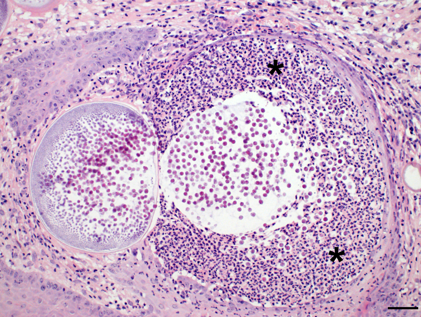
Histopathology at this stage shows:
- Thick-walled sporangia in various sizes
- Central sporangial cavity with developing endospores
- Surrounding granulomatous inflammation
3. Sporangial Rupture (Endospore Release)
- Upon full maturation, the sporangium ruptures, releasing numerous endospores into the surrounding tissue.
- Rupture is likely caused by:
◦ Pressure from accumulated endospores
◦ Host immune reaction
◦ Natural lifecycle progression
The released endospores can:
◦ Invade adjacent tissue
◦ Initiate new trophic forms
◦ Maintain the cycle locally, without needing a systemic or vector-based spread
DISCLAIMER-Some patients go to net and directly take treatment from there which can lead to catastrophic consequences-Then- Many people ask then why to read all this text -the reason is that it helps you to understand the pathology better ,you can cooperate with treatment better ,your treating physician is already busy with his patients and he does not have sufficient time to explain you all the things right from ABCD ,so it is always better to have some knowledge of the disease /disorder you are suffering from.
Immune response to released endospores includes:
- Lymphocytes
- Plasma cells
- Neutrophils
- Multinucleated giant cells, attempting to engulf sporangia or endospores
- Granuloma formation
This immune response contributes to tissue fibrosis, polyp formation, and chronicity of the lesion.
Modes of Reproduction
Rhinosporidium seeberi reproduces asexually via:
- Endosporogenesis: Formation of endospores within a sporangium
- There is no evidence of sexual reproduction, although genomic studies may eventually clarify this.

Environmental Stage (Hypothetical)
Though not yet demonstrated, many researchers hypothesize an external (aquatic) environmental stage:
- The organism may survive and multiply in water and mud, especially in stagnant ponds.
- Zoospores or immature forms may exist in water, waiting to infect a host.
- Some researchers suggest the possibility of aquatic vectors or intermediate hosts such as fish or amphibians, although this remains unproven.
Why In Vitro Culture Is Not Possible (Yet)
Rhinosporidium seeberi has never been cultured successfully in artificial media, despite extensive efforts.
Reasons include:
- Strict host-dependence
- Complex developmental cycle
- Unique nutritional requirements
- Obligate parasitism
This limitation significantly hinders research and contributes to our incomplete understanding of its life cycle.
Host Response and Chronicity
The chronic nature of rhinosporidiosis is due in part to:
- Continuous endospore release and re-infection
- Immune evasion by sporangial wall
- Failure of host to clear organism entirely
- Incomplete surgical excision leads to frequent recurrences
Comparison with Other Protists
The life cycle of R. seeberi resembles that of other Mesomycetozoea, such as:
- Dermocystidium spp. (fish pathogen)
- Ichthyophonus spp.
- These also form spherical sporangia and internal endospores, and may exist in aquatic environments.
Clinical Relevance of Life Cycle Understanding
Understanding the life cycle of R. seeberi has direct implications in:
1. Diagnosis
- Recognizing various developmental stages histologically (trophic forms, immature, and mature sporangia)
- Use of special stains like PAS and GMS for visualization
2. Treatment
- Surgical excision with cauterization of base is essential to eliminate local endospores
- Medical therapy with antifungals or antibiotics is ineffective
3. Prevention
- Avoidance of bathing in stagnant water in endemic areas
- Personal hygiene in rural or tropical regions
4. Research
- Molecular analysis of life cycle stages could help develop targeted therapies or vaccines
- Environmental DNA detection may offer new diagnostic or preventive tools
Conclusion
The life cycle of Rhinosporidium seeberi is a fascinating and complex process that is entirely host-bound, involving:
- A trophic phase,
- Development into a mature sporangium, and
- Rupture to release endospores.
Despite over a century of study, our understanding remains limited due to its in vitro unculturability and unique phylogenetic position. Continued research using molecular and genomic tools is essential for unlocking the full biology of this enigmatic pathogen.
FOR FURTHER INFORMATION IN GREAT DETAIL ON Rhinosporidiosis PL CLICK ON THE LINK GIVEN BELOW-It is always better to view links from laptop/desktop rather than mobile phone as they may not be seen from mobile phone. ,in case of technical difficulties you need to copy paste this link in google search. In case if you are viewing this blog from mobile phone you need to click on the three dots on the right upper corner of your mobile screen and ENABLE DESKTOP VERSION.
FOR INFORMATION IN GREAT DETAIL ON Rhinosporidiosis Pathology Outline PL CLICK ON THE LINK GIVEN BELOW-It is always better to view links from laptop/desktop rather than mobile phone as they may not be seen from mobile phone. ,in case of technical difficulties you need to copy paste this link in google search. In case if you are viewing this blog from mobile phone you need to click on the three dots on the right upper corner of your mobile screen and ENABLE DESKTOP VERSION.
FOR INFORMATION IN GREAT DETAIL ON Rhinosporidiosis Treatment PL CLICK ON THE LINK GIVEN BELOW-It is always better to view links from laptop/desktop rather than mobile phone as they may not be seen from mobile phone. ,in case of technical difficulties you need to copy paste this link in google search. In case if you are viewing this blog from mobile phone you need to click on the three dots on the right upper corner of your mobile screen and ENABLE DESKTOP VERSION.
FOR INFORMATION IN GREAT DETAIL ON Rhinosporidiosis Symptoms PL CLICK ON THE LINK GIVEN BELOW-It is always better to view links from laptop/desktop rather than mobile phone as they may not be seen from mobile phone. ,in case of technical difficulties you need to copy paste this link in google search. In case if you are viewing this blog from mobile phone you need to click on the three dots on the right upper corner of your mobile screen and ENABLE DESKTOP VERSION.
FOR FURTHER INFORMATION IN GREAT DETAIL ON Rhinosporidiosis Histology PL CLICK ON THE LINK GIVEN BELOW-It is always better to view links from laptop/desktop rather than mobile phone as they may not be seen from mobile phone. ,in case of technical difficulties you need to copy paste this link in google search. In case if you are viewing this blog from mobile phone you need to click on the three dots on the right upper corner of your mobile screen and ENABLE DESKTOP VERSION.
If any patient has any ENT -Ear nose throat problems and requires any , consultation ,online consultation ,or surgery in clinic of ENT specialist Doctor Dr Sagar Rajkuwar ,he may TAKE APPOINTMENT BY CLICKING ON THE LINK GIVEN BELOW-
Clinic address of ENT SPECIALIST doctor Dr Sagar Rajkuwar-
Prabha ENT clinic, plot no 345,Saigram colony, opposite Indoline furniture Ambad link road ,Ambad ,1 km from Pathardi phata Nashik ,422010 ,Maharashtra, India-Dr Sagar Rajkuwar (MS-ENT), Cel no- 7387590194 , 9892596635