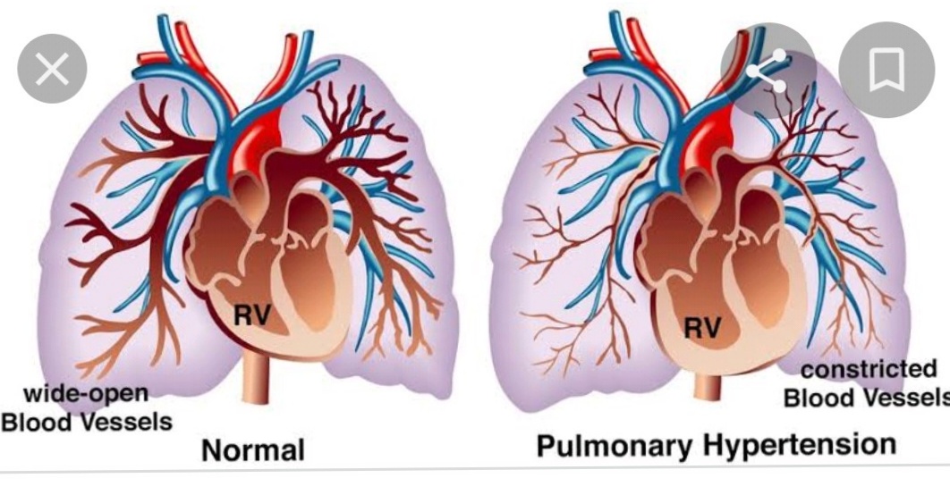
Pulmonary Hypertension Complications-Various aspects-
Pulmonary hypertension (PH) may result in a number of complications, such as heart failure, blood clots, and lung bleeding. These complications can pose a serious threat to life.
Heart complications
Right-sided heart failure: Also referred to as cor pulmonale, this condition arises when the right lower chamber of the heart enlarges and becomes weak.
Arrhythmias: Irregular heartbeats that may be life-threatening.
Pericardial effusion: Accumulation of fluid surrounding the heart.
Blood clot complications
Blood clots in the lungs: PH raises the likelihood of blood clots occurring in the small arteries of the lungs.
Bleeding complications
Bleeding in the lungs: PH can result in life-threatening bleeding within the lungs, potentially leading to coughing up blood.
Pulmonary artery aneurysm: A rupture in the pulmonary artery may lead to hemorrhage.
Pregnancy complications
Cardiac arrest: PH may elevate the risk of cardiac arrest during pregnancy.
Heart attack: PH may raise the risk of heart attack during pregnancy.
Respiratory failure: PH can heighten the risk of respiratory failure during pregnancy.
Stroke: PH may increase the likelihood of stroke occurring during pregnancy.
Other complications include:
Anemia
Liver damage
Bundle branch blocks
Syncope, or temporary loss of consciousness
PH is a condition that deteriorates over time and can be deadly if not treated. Treatments can assist in alleviating symptoms and managing the condition.


1. Extrinsic compression of the left main stem coronary artery in a 37-year-old female with pulmonary hypertension presenting typical angina: (a) An electrocardiogram-gated coronal CT is showing extrinsic compression of the left main stem coronary artery (black block arrow) caused by the aneurysmal main pulmonary artery (MPA). (b) The axial CT image of the same patient is revealing a sinus venosus-type atrial septal defect (white block arrow) along with a dilated right ventricle (RV). LA, left atrium; RA, right atrium.
Extrinsic compression of the proximal airways due to pulmonary artery dilatation is frequently observed in congenital heart disease. This can result in asthma-like symptoms such as wheezing and coughing, and it is not likely to improve with standard treatment. Furthermore, a similar phenomenon may occur at the level of the distal small airways, due to their closeness to the distal pulmonary arteries. It remains uncertain whether this is solely a mechanical issue, or if there is an involvement of an imbalance between vasoconstrictive and bronchoconstrictive agents.
2. Extrinsic compression of the airways in a 27-year-old woman with pulmonary hypertension and increasing dyspnoea: compression of the right main bronchus (block arrows) by the enlarged right pulmonary artery (RPA) on mediastinal (left) and lung window (right).
Pulmonary artery dissection
Pulmonary artery dissection is an uncommon and potentially devastating complication, most of which are identified only post-mortem. It is linked with congenital heart disease and may arise idiopathically from chronic PH or iatrogenically from catheterization. The pulmonary artery dissection generally ruptures into the pericardium resulting in tamponade, rather than progressing further downstream with a re-entrance site. The most frequent site of dissection is the main pulmonary artery. On CT, its shape may resemble that of a typical aortic dissection, featuring an intimal flap and increased contrast attenuation within the true lumen. There have been case reports in which this is the initial indication of chronically undiagnosed PH. Symptoms are vague and vary from chest pain to dyspnoea.
3. Pulmonary artery dissection affecting the left pulmonary artery (arrow). Additionally, there is a brief film accessible online.
Alveolar haemorrhage
Alveolar haemorrhage in individuals with PAH is multifactorial and might result from anticoagulation therapy and a continuous intravenous infusion of pulmonary vasodilator medications. There exists a debate regarding the use of anticoagulants in patients with PH, and a recent evaluation utilizing the European PH registry Comparative, Prospective Registry of Newly Initiated Therapies for Pulmonary Hypertension supports anticoagulation for patients with idiopathic PAH, but not for other subgroups.
Haemorrhage can occur from a rupture of a pulmonary artery aneurysm, which, when present, primarily exists within the main pulmonary artery. It may result in ground glass and consolidative alterations on CT and dynamic extravasation of contrast during an invasive angiogram . Cabezas’ group researched a large population of patients with Group 1 PH, and the incidence of pulmonary artery aneurysm was found to be 12. 2%. Previously, this was identified only at necropsy, but it is increasingly recognized due to the rising application of cross-sectional imaging. The management of pulmonary artery aneurysm, whether through surgery or medical optimization, is contentious in the literature.
4. Alveolar haemorrhage in a 45-year-old woman with pulmonary hypertension following right heart catheterization: (a) coronal maximum intensity projection of an MR pulmonary angiogram shows a proximal blockage of the right middle and lower lobes (black block arrow) and proximal webs in the left lower lobe (thin white arrows) consistent with chronic thromboembolic disease. (b) The patient experienced haemoptysis after the right heart catheterization. The axial CT image illustrates a pseudoaneurysm of the posterior basal left lower lobe artery (white block arrow) and adjacent alveolar haemorrhage that occurred due to the forced movement of the catheter across the intraluminal webs. (c) Selected images from catheter pulmonary angiography before (left) and after (right) embolization of the left lower lobe pseudoaneurysm (block white arrows).
In situ thrombosis
In situ thrombosis refers to the formation of thrombus within the pulmonary arteries, without embolism originating from a distal deep-vein thrombosis. Thrombus formation is frequently seen in severe hypertension due to various causes. The proposed mechanisms differ, and this may arise from a mix of wall shear stress from turbulent flow, vascular dilation leading to stasis, and vascular endothelial injury in PH. It might also result from increased thrombin activity and disruption of the thrombolytic pathway. In situ thrombosis is prevalent in congenital heart disease, with up to one-fifth of patients with Eisenmenger’s syndrome potentially having pulmonary thrombi. These thrombi can calcify and might be part of a partially thrombosed pulmonary artery. Unlike acute thrombi, in situ thrombi are positioned peripherally with lobulated shapes and generally form an obtuse angle with the vessel wall. They may additionally exhibit coarse calcification. Interestingly, patients with Eisenmenger’s syndrome have abnormal coagulation and are at risk of haemorrhage. This complicates the management of their treatment.
5. In situ thrombus in a 57-year-old woman with longstanding pulmonary hypertension due to congenital heart disease: (a) the axial non-electrocardiogram-gated CT pulmonary angiogram shows calcified thrombus lining the proximal pulmonary arteries (white block arrows). (b) There is dilation of the right atrium (RA) and right ventricle (RV), with right ventricular hypertrophy and an ostium secundum atrial septal defect (black thin arrow). The segmental vessels are enlarged and contain partially occlusive calcified thrombus.
Balloon angioplasty complications
The history of balloon pulmonary angioplasty (BPA) as a method of treating chronic thromboembolic pulmonary hypertension (CTEPH) goes back to 1983. The standard treatment is pulmonary endarterectomy (PEA), which shows immediate post-operative improvements in hemodynamics and enhanced survival rates. BPA may serve as an alternative for patients who are not candidates for surgery. Research has indicated that there is no immediate improvement in hemodynamics, but follow-up reveals a reduction in right atrial pressure and pulmonary arterial pressure, along with an increase in cardiac output. BPA can address distal lesions that PEA typically cannot reach, primarily the segmental and subsegmental pulmonary arteries. However, these lesions remain under significant pressure, and patients with severely compromised baseline hemodynamics are susceptible to reperfusion edema. The reported incidence of this complication is 53–61%. A potentially life-threatening complication is pulmonary artery perforation, leading to hemorrhage and exsanguination, which is less prevalent and occurs in up to 7% of cases.
Patients who have had BPA continue to receive pulmonary hypertension (PH)-specific treatment, and it remains to be seen whether this influences the improvement in hemodynamics. Additional research and long-term data are necessary to assess the effectiveness of BPA as an alternative to PEA.
Cardiac complications
Right heart failure
As pulmonary artery resistance and pressure escalate, the right ventricle initially responds to this pressure overload with hypertrophy and eventually dilates to enhance preload and sustain stroke volume. Over time, the right ventricle can no longer adapt, although the exact mechanism remains unclear. This may lead to functional tricuspid regurgitation, pericardial effusions, and cardiac cirrhosis.


