
Rhinoscleroma vs Rhinosporidiosis-various aspects-
Despite being rare, granulomatous illnesses that impact the nasal passages and upper respiratory tract, rhinoscleroma and rhinosporidiosis have unique features and are caused by different pathogens. The Klebsiella rhinoscleromatis bacteria causes rhinoscleroma, whereas the Rhinosporidium seeberi fungus causes rhinosporidiosis.
![]()
Rhinoscleroma:
Causative factor:
A bacterium called Klebsiella rhinoscleromatis.
Transmission:
Mainly through direct contact with tainted objects or sick droplets.
Symptoms:
May manifest as nasal congestion, a runny nose, and, in the later stages, as stenosis (narrowing) and scarring of the nasal passages and larynx.
Histology:
Russell bodies, or immunoglobulin deposits, and Mikulicz cells, or foamy macrophages, are its defining characteristics.
Treatment:
Usually includes extended antibiotic treatment with drugs like ciprofloxacin and doxycycline.
Geographical Distribution:
Although it is prevalent in some tropical and subtropical areas, it may also be found among immigrant communities in other locations.
If Any Patient of ENT Requires Any Surgery, Opd Consultation Or Online Consultation In Clinic of ENT Specialist Doctor Dr. Sagar Rajkuwar ,He May Contact Him At The Following Address-
Prabha ENT Clinic, Plot no 345,Saigram Colony, Opposite Indoline Furniture Ambad Link Road ,Ambad ,1 km From Pathardi Phata Nashik ,422010 ,Maharashtra, India-Dr. Sagar Rajkuwar (MS-ENT), Cell No- 7387590194, 9892596635
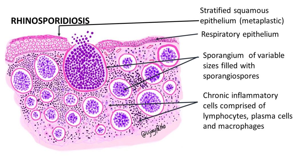
Rhinosporidiosis:
- Cause: a fungus called Rhinosporidium seeberi.
- Transmission: Infection usually results from coming into touch with the fungus, frequently through ontaminated water or soil.
- Symptoms: Marked by the development of brittle, polypoid masses (polyps) in the eyes, nose, and oropharynx. These growths are frequently reddish and bleed easily.
- Histology: Demonstrates the existence of R. seeberi sporangia, which are spore-containing structures.
- Treatment: The main course of treatment is surgical removal of the polyps, and dapsone may be used to avoid a relapse.
- Geographic Distribution: Mostly found in India and Sri Lanka.
Some disorders, such as inflammatory illnesses, infectious diseases, tumors, trauma, and cocaine usage, can lead to granulomatous diseases of the nose. Regular examinations, medicinal therapy, and sometimes surgical management are necessary for certain illnesses, such as invasive fungal rhinosinusitis and rhinoscleroma, that follow a chronic course.
For Update On Further Important Health Related Topics And Frequently Asked Questions On Health Topics By General Population Please Click On The Link Given Below To Join Our WhatsApp Group –
https://chat.whatsapp.com/Lv3NbcguOBS5ow6X9DpMMA
Rhinoscleroma
- It’s a granulomatous bacterial infection.
- The cause of this illness is the gram-negative bacterium Klebsiella rhinoscleromatis.
- In India, rhinoscleroma is more common in the north than in the south.
- Through airborne droplets, the virus spreads.
- The clinical manifestations are categorized into several stages:
1. Stage 1: Atrophic stage
During this phase, the bacteria kill the cells that produce mucin. These cells contain immunoglobulins, and because they are destroyed, there will be no immunoglobulins, which will allow the bacteria to flourish there and prevent the formation of mucus.
Dryness of the nose causes crust and foul-smelling purulent nasal discharge.
2. Step 2: Granulomatous Phase
The granuloma is formed during this phase as the body’s immune system begins to combat the bacteria.
A woody sensation is caused by nodules in the nose.
This causes the formation of a hard, woody nodule that deforms the nose, often referred to as a Hebra nose.
The nodules are located in the lower part of the outer lip and nose.
o The nodules are occasionally nonulcerative and pain-free.
3. Stage 3: Cicatrizing stage or stage of healing – The third stage of rhinoscleroma is also known as the stage of the healing period.
◦ The nasal and nasopharynx are constricted during this phase. Additionally, it is referred to as a Tapir nose deformity.
◦ The disease begins in the nose and spreads to the pharynx, larynx, trachea, and bronchus.
The subglottic larynx is the area most severely affected by laryngeal scleroma.
Rhinoscleroma diagnosis
- Own biopsy or histopathological examination there are two types of cells present. These are:
- Mikulicz cells: These are histiocytes with vacuoles.
o Russell bodies – These are eosinophilic plasma cell inclusions. - MacConkey’s agar medium is the culture medium that is used.
DISCLAIMER-Some patients go to net and directly take treatment from there which can lead to catastrophic consequences-Then- Many people ask then why to read all this text -the reason is that it helps you to understand the pathology better ,you can cooperate with treatment better ,your treating physician is already busy with his patients and he does not have sufficient time to explain you all the things right from ABCD ,so it is always better to have some knowledge of the disease /disorder you are suffering from.
Rhinoscleroma Therapy
- The first line of treatment for less severe illness is medical therapy.
For at least four to six weeks, use tetracycline and streptomycin. - The culture samples are repeated and analyzed four to six weeks after the start of treatment.
If the culture is positive, we should continue the therapy, but if it is negative, we must conduct another repeat culture after a week. The treatment should be stopped if the culture is still negative after a week. - During the cicatrization phase, steroids are administered to lessen fibrosis.
- The skin irritation is treated with acriflavine. It is a local treatment for the nodules.
- Surgery is the best option for debriding necrotic tissue if there is a high concentration of it.
Irradiation is occasionally used.

Rhinosporidiosis
- It’s neither a bacterial, viral, nor fungal infection. The aquatic protozoa Rhinosporidium Seeberi is the cause of this illness.
- It’s a disease that spreads via water.
- It is common in South Asian nations such as India, Pakistan, and Sri Lanka.
Rhinosporidiosis’s Pathogenesis
- The nasal cavity’s epidermis is where these protozoa reproduce in layers, producing a red polypoidal mass.
- Spores that resemble white spots are found in the mass, which is very vascular.
- The nasal cavity contains a strawberry-like mass that is similar to this red polypoidal mass with white patches.
On histopathological examination:
- The sporangia with the spores will be found.
- Diagnosis cannot be established on culture.
- It is mainly established by history, clinical examination, and histopathological examination.
Symptoms of Rhinosporidiosis
The bleeding from the nose is the primary manifestation of rhinosporidiosis.
- The patient will present with features of nasal obstruction and difficulty in breathing.
- External polypoidal nose results in cosmetic problems and the patient’s routine social life will be altered.
Treatment of Rhinosporidiosis
- Surgery followed by medical treatment is the intervention of choice.
- Complete excision of the mass with a diathermy knife and cauterization of its base is done.
- Recurrence is very common in rhinosporidiosis.
- Medical treatment is Dapsone. However, Dapsone is partially effective and can be given to reduce the recurrence.
Major Variations:
The Cause:
Rhinosporidiosis (fungal) vs. Rhinoscleroma (bacterial).
Features of the Lesion:
While rhinosporidiosis is distinguished by polypoid development, rhinoscleroma can result in scarring and stenosis.
Method of Treatment:
Treatment for Rhinoscleroma using antibiotics vs. surgical excision and antifungal drugs for Rhinosporidiosis.
Endemic Regions:
Rhinosporidiosis is more common in India and Sri Lanka, whereas rhinoscleroma is more widely distributed throughout tropical and subtropical climates.
FOR FURTHER INFORMATION IN GREAT DETAIL ON Rhinosporidiosis PL CLICK ON THE LINK GIVEN BELOW-It is always better to view links from laptop/desktop rather than mobile phone as they may not be seen from mobile phone. ,in case of technical difficulties you need to copy paste this link in google search. In case if you are viewing this blog from mobile phone you need to click on the three dots on the right upper corner of your mobile screen and ENABLE DESKTOP VERSION.
FOR INFORMATION IN GREAT DETAIL ON Rhinosporidiosis Pathology Outline PL CLICK ON THE LINK GIVEN BELOW-It is always better to view links from laptop/desktop rather than mobile phone as they may not be seen from mobile phone. ,in case of technical difficulties you need to copy paste this link in google search. In case if you are viewing this blog from mobile phone you need to click on the three dots on the right upper corner of your mobile screen and ENABLE DESKTOP VERSION.
FOR INFORMATION IN GREAT DETAIL ON Rhinosporidiosis Treatment PL CLICK ON THE LINK GIVEN BELOW-It is always better to view links from laptop/desktop rather than mobile phone as they may not be seen from mobile phone. ,in case of technical difficulties you need to copy paste this link in google search. In case if you are viewing this blog from mobile phone you need to click on the three dots on the right upper corner of your mobile screen and ENABLE DESKTOP VERSION.
FOR INFORMATION IN GREAT DETAIL ON Rhinosporidiosis Symptoms PL CLICK ON THE LINK GIVEN BELOW-It is always better to view links from laptop/desktop rather than mobile phone as they may not be seen from mobile phone. ,in case of technical difficulties you need to copy paste this link in google search. In case if you are viewing this blog from mobile phone you need to click on the three dots on the right upper corner of your mobile screen and ENABLE DESKTOP VERSION.
FOR FURTHER INFORMATION IN GREAT DETAIL ON Rhinosporidiosis Histology PL CLICK ON THE LINK GIVEN BELOW-It is always better to view links from laptop/desktop rather than mobile phone as they may not be seen from mobile phone. ,in case of technical difficulties you need to copy paste this link in google search. In case if you are viewing this blog from mobile phone you need to click on the three dots on the right upper corner of your mobile screen and ENABLE DESKTOP VERSION.
FOR FURTHER INFORMATION IN GREAT DETAIL ON Rhinosporidiosis Life Cycle PL CLICK ON THE LINK GIVEN BELOW-It is always better to view links from laptop/desktop rather than mobile phone as they may not be seen from mobile phone. ,in case of technical difficulties you need to copy paste this link in google search. In case if you are viewing this blog from mobile phone you need to click on the three dots on the right upper corner of your mobile screen and ENABLE DESKTOP VERSION.
If any patient has any ENT -Ear nose throat problems and requires any , consultation ,online consultation ,or surgery in clinic of ENT specialist Doctor Dr Sagar Rajkuwar ,he may TAKE APPOINTMENT BY CLICKING ON THE LINK GIVEN BELOW-
Clinic address of ENT SPECIALIST doctor Dr Sagar Rajkuwar-
Prabha ENT clinic, plot no 345,Saigram colony, opposite Indoline furniture Ambad link road ,Ambad ,1 km from Pathardi phata Nashik ,422010 ,Maharashtra, India-Dr Sagar Rajkuwar (MS-ENT), Cel no- 7387590194 , 9892596635


